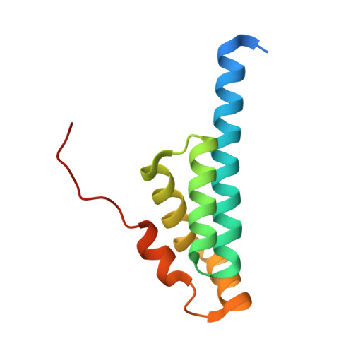
1UTU

Crystal structure of novel protein EMSY truncate


Experimental Data Snapshot

  • Method: X-RAY DIFFRACTION
  • Resolution: 2.00 Å
  • R-Value Free: 0.272 
  • R-Value Work: 0.207 
  • R-Value Observed: 0.207 

wwPDB Validation   3D Report Full Report


This is version 1.4 of the entry. See complete history


Literature

Binding of Emsy to Hp1Beta: Implications for Recruitment of Hp1Beta and Bs69.

Ekblad, C.M.Chavali, G.B.Basu, B.P.Freund, S.M.Veprintsev, D.Hughes-Davies, L.Kouzarides, T.Doherty, A.J.Itzhaki, L.S.

(2005) EMBO Rep 6: 675

  • DOI: https://doi.org/10.1038/sj.embor.7400415
  • Primary Citation of Related Structures:  
    1UTU

  • PubMed Abstract: 

    EMSY is a large nuclear protein that binds to the transactivation domain of BRCA2. EMSY contains an approximately 100-residue segment at the amino terminus called the ENT (EMSY N-terminal) domain. Plant proteins containing ENT domains also contain members of the royal family of chromatin-remodelling domains. It has been proposed that EMSY may have a role in chromatin-related processes. This is supported by the observation that a number of chromatin-regulator proteins, including HP1beta and BS69, bind directly to EMSY by means of a conserved motif adjacent to the ENT domain. Here, we report the crystal structure of residues 1-108 of EMSY at 2.0 A resolution. The structure contains both the ENT domain and the HP1beta/BS69-binding motif. This binding motif forms an extended peptide-like conformation that adopts distinct orientations in each subunit of the dimer. Biophysical and nuclear magnetic resonance analyses show that the main complex formed by EMSY and the chromoshadow domain of HP1 (HP1-CSD) consists of one EMSY dimer sandwiched between two HP1-CSD dimers. The HP1beta-binding motif is necessary and sufficient for EMSY to bind to the chromoshadow domain of HP1beta.


  • Organizational Affiliation

    MRC Cancer Cell Unit, Hutchison/MRC Research Centre, Hills Road, Cambridge CB2 2XZ, UK.


Macromolecules
Find similar proteins by:  (by identity cutoff)  |  3D Structure
Entity ID: 1
MoleculeChains Sequence LengthOrganismDetailsImage
EMSY
A, B
108Homo sapiensMutation(s): 0 
UniProt & NIH Common Fund Data Resources
Find proteins for Q7Z589 (Homo sapiens)
Explore Q7Z589 
Go to UniProtKB:  Q7Z589
PHAROS:  Q7Z589
GTEx:  ENSG00000158636 
Entity Groups  
Sequence Clusters30% Identity50% Identity70% Identity90% Identity95% Identity100% Identity
UniProt GroupQ7Z589
Sequence Annotations
Expand
  • Reference Sequence
Experimental Data & Validation

Experimental Data

  • Method: X-RAY DIFFRACTION
  • Resolution: 2.00 Å
  • R-Value Free: 0.272 
  • R-Value Work: 0.207 
  • R-Value Observed: 0.207 
  • Space Group: P 1 21 1
Unit Cell:
Length ( Å )Angle ( ˚ )
a = 51.178α = 90
b = 32.642β = 92.51
c = 51.607γ = 90
Software Package:
Software NamePurpose
CNSrefinement
MOSFLMdata reduction
SCALAdata scaling
MOLREPphasing

Structure Validation

View Full Validation Report


Entry History 

Deposition Data

Revision History  (Full details and data files)

  • Version 1.0: 2005-10-13
    Type: Initial release
  • Version 1.1: 2011-05-08
    Changes: Version format compliance
  • Version 1.2: 2011-07-13
    Changes: Version format compliance
  • Version 1.3: 2017-07-05
    Changes: Data collection
  • Version 1.4: 2024-05-08
    Changes: Data collection, Database references, Other