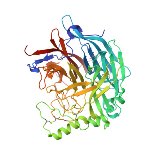
1USR

Newcastle disease virus hemagglutinin-neuraminidase: Evidence for a second sialic acid binding site and implications for fusion


Experimental Data Snapshot

  • Method: X-RAY DIFFRACTION
  • Resolution: 2.00 Å
  • R-Value Free: 0.226 
  • R-Value Work: 0.190 
  • R-Value Observed: 0.190 

wwPDB Validation   3D Report Full Report


This is version 3.0 of the entry. See complete history


Literature

Second Sialic Acid Binding Site in Newcastle Disease Virus Hemagglutinin-Neuraminidase: Implications for Fusion

Zaitsev, V.Von Itzstein, M.Groves, D.Kiefel, M.Takimoto, T.Portner, A.Taylor, G.

(2004) J Virol 78: 3733

  • DOI: https://doi.org/10.1128/jvi.78.7.3733-3741.2004
  • Primary Citation of Related Structures:  
    1USR, 1USX

  • PubMed Abstract: 
  • Paramyxoviruses are the leading cause of respiratory disease in children. Several paramyxoviruses possess a surface glycoprotein, the hemagglutinin-neuraminidase (HN), that is involved in attachment to sialic acid receptors, promotion of fusion, and removal of sialic acid from infected cells and progeny virions ...

    Paramyxoviruses are the leading cause of respiratory disease in children. Several paramyxoviruses possess a surface glycoprotein, the hemagglutinin-neuraminidase (HN), that is involved in attachment to sialic acid receptors, promotion of fusion, and removal of sialic acid from infected cells and progeny virions. Previously we showed that Newcastle disease virus (NDV) HN contained a pliable sialic acid recognition site that could take two states, a binding state and a catalytic state. Here we present evidence for a second sialic acid binding site at the dimer interface of HN and present a model for its involvement in cell fusion. Three different crystal forms of NDV HN now reveal identical tetrameric arrangements of HN monomers, perhaps indicative of the tetramer association found on the viral surface.


    Related Citations: 
    • Crystal Structure of the Multifunctional Paramyxovirus Hemagglutinin-Neuraminidase
      Crennell, S., Takimoto, T., Portner, A., Taylor, G.
      (2000) Nat Struct Biol 7: 1068
    • Crystallization of Newcastle Disease Virus Hemagglutinin-Neuraminidase Glycoprotein
      Takimoto, T., Taylor, G.L., Crennell, S.J., Scroggs, R.A., Portner, A.
      (2000) Virology 270: 208

    Organizational Affiliation

    Centre for Biomolecular Sciences, University of St. Andrews, St. Andrews, Fife KY16 9ST, United Kingdom.


Macromolecules
Find similar proteins by:  (by identity cutoff)  |  3D Structure
Entity ID: 1
MoleculeChainsSequence LengthOrganismDetailsImage
HEMAGGLUTININ-NEURAMINIDASE GLYCOPROTEINA, B454Avian orthoavulavirus 1Mutation(s): 0 
EC: 3.2.1.18
UniProt
Find proteins for P32884 (Newcastle disease virus (strain Beaudette C/45))
Explore P32884 
Go to UniProtKB:  P32884
Entity Groups  
Sequence Clusters30% Identity50% Identity70% Identity90% Identity95% Identity100% Identity
UniProt GroupP32884
Protein Feature View
Expand
  • Reference Sequence
Oligosaccharides

Help

Entity ID: 2
MoleculeChainsChain Length2D DiagramGlycosylation3D Interactions
N-acetyl-alpha-neuraminic acid-(2-6)-methyl 6-thio-beta-D-galactopyranosideC 2N/AN/A Oligosaccharides Interaction
Glycosylation Resources
GlyTouCan:  G18879GM
GlyCosmos:  G18879GM
Small Molecules
Ligands 4 Unique
IDChainsName / Formula / InChI Key2D Diagram3D Interactions
SIA
Query on SIA

Download Ideal Coordinates CCD File 
I [auth B]N-acetyl-alpha-neuraminic acid
C11 H19 N O9
SQVRNKJHWKZAKO-YRMXFSIDSA-N
 Ligand Interaction
DAN
Query on DAN

Download Ideal Coordinates CCD File 
D [auth A],
G [auth B]
2-DEOXY-2,3-DEHYDRO-N-ACETYL-NEURAMINIC ACID
C11 H17 N O8
JINJZWSZQKHCIP-UFGQHTETSA-N
 Ligand Interaction
NDG
Query on NDG

Download Ideal Coordinates CCD File 
E [auth A],
H [auth B]
2-acetamido-2-deoxy-alpha-D-glucopyranose
C8 H15 N O6
OVRNDRQMDRJTHS-PVFLNQBWSA-N
 Ligand Interaction
CA
Query on CA

Download Ideal Coordinates CCD File 
F [auth A],
J [auth B]
CALCIUM ION
Ca
BHPQYMZQTOCNFJ-UHFFFAOYSA-N
 Ligand Interaction
Experimental Data & Validation

Experimental Data

  • Method: X-RAY DIFFRACTION
  • Resolution: 2.00 Å
  • R-Value Free: 0.226 
  • R-Value Work: 0.190 
  • R-Value Observed: 0.190 
  • Space Group: P 21 21 2
Unit Cell:
Length ( Å )Angle ( ˚ )
a = 175.48α = 90
b = 99.26β = 90
c = 64.33γ = 90
Software Package:
Software NamePurpose
CNSrefinement
DENZOdata reduction
SCALEPACKdata scaling
CNSphasing

Structure Validation

View Full Validation Report


Entry History 

Deposition Data

Revision History  (Full details and data files)

  • Version 1.0: 2004-01-02
    Type: Initial release
  • Version 1.1: 2011-05-08
    Changes: Version format compliance
  • Version 1.2: 2011-07-13
    Changes: Version format compliance
  • Version 2.0: 2020-07-01
    Changes: Atomic model, Data collection, Derived calculations, Non-polymer description, Structure summary
  • Version 3.0: 2020-07-29
    Type: Remediation
    Reason: Carbohydrate remediation
    Changes: Atomic model, Data collection, Derived calculations, Structure summary