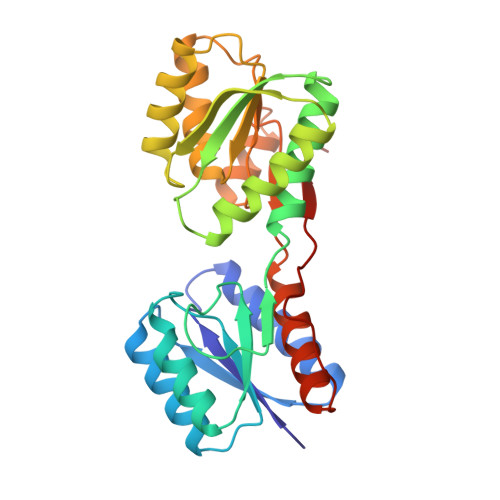
Multiple open forms of ribose-binding protein trace the path of its conformational change.
Bjorkman, A.J., Mowbray, S.L.(1998) J Mol Biology 279: 651-664
- PubMed: 9641984 Search on PubMed
- DOI: https://doi.org/10.1006/jmbi.1998.1785
- Primary Citation Related Structures:
1BA2, 1URP - PubMed Abstract:
Conformational changes are necessary for the function of bacterial periplasmic receptors in chemotaxis and transport. Such changes allow entry and exit of ligand, and enable the correct interaction of the ligand-bound proteins with the membrane components of each system. Three open, ligand-free forms of the Escherichia coli ribose-binding protein were observed here by X-ray crystallographic studies. They are opened by 43 degrees, 50 degrees and 64 degrees with respect to the ligand-bound protein reported previously. The three open forms are not distinct, but show a clear relationship to each other. All are the product of a similar opening motion, and are stabilized by a new, almost identical packing interface between the domains. The changes are generated by similar bond rotations, although some differences in the three hinge segments are needed to accommodate the various structural scenarios. Some local repacking also occurs as interdomain contacts are lost. The least open (43 degrees) form is probably the dominant one in solution under normal conditions, although a mixture of species seems likely. The open and closed forms have distinct surfaces in the regions known to be important in chemotaxis and transport, which will differentiate their interactions with the membrane components. It seems certain that the conformational path that links the forms described here is that followed during ligand retrieval, and in ligand release into the membrane-bound permease system.
- Department of Molecular Biology, Swedish Agricultural University, Uppsala, Sweden.
Organizational Affiliation: