1URL

N-TERMINAL DOMAIN OF SIALOADHESIN (MOUSE) IN COMPLEX WITH GLYCOPEPTIDE


Experimental Data Snapshot

  • Method: X-RAY DIFFRACTION
  • Resolution: 2.40 Å
  • R-Value Free: 0.273 
  • R-Value Work: 0.225 
  • R-Value Observed: 0.225 

wwPDB Validation   3D Report Full Report


Ligand Structure Quality Assessment 


This is version 1.5 of the entry. See complete history


Literature

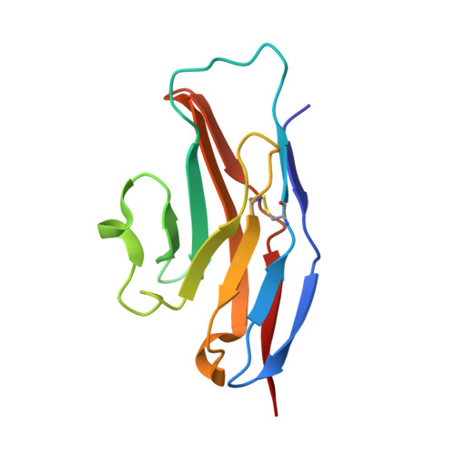
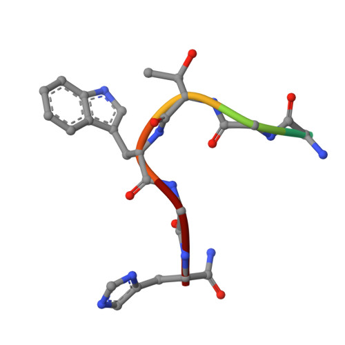
Complex of Sialoadhesin with a Glycopeptide Ligand

Bukrinsky, J.T.Hilaire, P.M.S.Meldal, M.Crocker, P.R.Henriksen, A.

(2004) Biochim Biophys Acta 1702: 173

  • DOI: https://doi.org/10.1016/j.bbapap.2004.08.015
  • Primary Citation of Related Structures:  
    1URL

  • PubMed Abstract: 

    Sialoadhesin is a sialic acid-binding immunoglobulin-like lectin (Siglec), expressed on subsets of macrophages. It is a model system for Siglec receptor-mediated cell surface interactions through binding of sialylated glycoconjugates. The N-terminal sialoadhesin domain can mediate sialic acid-binding on its own. The structure of this domain has been determined in complex with a sialic acid-containing heptapeptide, (Ala-Gly-His-Thr(Neu5Ac)-Trp-Gly-His). The affinity of sialoadhesin for this ligand is four times higher than the affinity for the natural linkage 2,3'-sialyllactose. The structure of the glycopeptide complex suggests strategies for ligand optimization and provides possible explanations for the observed differences in specificities among the Siglecs.


  • Organizational Affiliation

    Department of Chemistry, The Carlsberg Laboratory, Gamle Carlsberg Vej 10, DK-2500 Valby, Denmark.


Macromolecules
Find similar proteins by:  (by identity cutoff)  |  3D Structure
Entity ID: 1
MoleculeChains Sequence LengthOrganismDetailsImage
SIALOADHESIN118Mus musculusMutation(s): 0 
UniProt & NIH Common Fund Data Resources
Find proteins for Q62230 (Mus musculus)
Explore Q62230 
Go to UniProtKB:  Q62230
IMPC:  MGI:99668
Entity Groups  
Sequence Clusters30% Identity50% Identity70% Identity90% Identity95% Identity100% Identity
UniProt GroupQ62230
Sequence Annotations
Expand
  • Reference Sequence

Find similar proteins by:  Sequence   |   3D Structure  

Entity ID: 2
MoleculeChains Sequence LengthOrganismDetailsImage
ALA-GLY-HIS-THR-TRP-GLY-HIA7synthetic constructMutation(s): 0 
Sequence Annotations
Expand
  • Reference Sequence
Small Molecules
Ligands 1 Unique
IDChains Name / Formula / InChI Key2D Diagram3D Interactions
SIA
Query on SIA

Download Ideal Coordinates CCD File 
C [auth B]N-acetyl-alpha-neuraminic acid
C11 H19 N O9
SQVRNKJHWKZAKO-YRMXFSIDSA-N
Modified Residues  1 Unique
IDChains TypeFormula2D DiagramParent
HIA
Query on HIA
B
L-PEPTIDE LINKINGC6 H10 N4 OHIS
Experimental Data & Validation

Experimental Data

  • Method: X-RAY DIFFRACTION
  • Resolution: 2.40 Å
  • R-Value Free: 0.273 
  • R-Value Work: 0.225 
  • R-Value Observed: 0.225 
  • Space Group: P 21 21 2
Unit Cell:
Length ( Å )Angle ( ˚ )
a = 42.23α = 90
b = 78.46β = 90
c = 34.22γ = 90
Software Package:
Software NamePurpose
CNSrefinement
MOSFLMdata reduction
SCALAdata scaling
AMoREphasing

Structure Validation

View Full Validation Report


Ligand Structure Quality Assessment 


Entry History 

Deposition Data

Revision History  (Full details and data files)

  • Version 1.0: 2004-10-20
    Type: Initial release
  • Version 1.1: 2011-05-08
    Changes: Version format compliance
  • Version 1.2: 2011-07-13
    Changes: Version format compliance
  • Version 1.3: 2018-01-17
    Changes: Data collection, Source and taxonomy
  • Version 1.4: 2020-07-29
    Type: Remediation
    Reason: Carbohydrate remediation
    Changes: Data collection, Derived calculations, Other, Structure summary
  • Version 1.5: 2023-12-13
    Changes: Data collection, Database references, Refinement description, Structure summary