1UPW | pdb_00001upw

Crystal structure of the human Liver X receptor beta ligand binding domain in complex with a synthetic agonist


Experimental Data Snapshot

  • Method: X-RAY DIFFRACTION
  • Resolution: 2.40 Å
  • R-Value Free: 
    0.295 (Depositor) 
  • R-Value Work: 
    0.250 (Depositor) 

Starting Model: experimental
View more details

wwPDB Validation 3D Report Full Report

Validation slider image for 1UPW

This is version 1.4 of the entry. See complete history

Literature

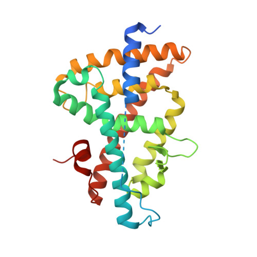
Crystal Structure of the Human Liver X Receptor Beta Ligand-Binding Domain in Complex with a Synthetic Agonist

Hoerer, S.Schmid, A.Heckel, A.Budzinski, R.M.Nar, H.

(2003) J Mol Biology 334: 853

  • DOI: https://doi.org/10.1016/j.jmb.2003.10.033
  • Primary Citation Related Structures: 
    1UPV, 1UPW

  • PubMed Abstract: 

    LXRbeta belongs to the nuclear hormone receptor superfamily of ligand-activated transcription factors. Its natural ligands are supposed to be oxidised derivatives of cholesterol. Stimulation of LXRbeta by agonists activates a number of genes that are involved in the regulation of lipid metabolism and cholesterol efflux from cells. Therefore, LXRbeta may represent a novel therapeutic target for the treatment of dyslipidemia and atherosclerosis.Here, we report the X-ray crystal structure of the LXRbeta ligand-binding domain in complex with a synthetic agonist, T-0901317. This compound occupies the ligand-binding pocket of the receptor, forms numerous lipophilic contacts with the protein and one crucial hydrogen bond to His435 and stabilises the agonist conformation of the receptor ligand-binding domain. The recruitment of the AF2-region of the protein is not achieved via direct polar interactions of the ligand with protein side-chains of this helical segment, but rather via few hydrophobic contacts and probably more importantly via indirect effects involving the pre-orientation of side-chains that surround the ligand-binding pocket and form the interface to the AF2-helix. On the basis of these results we propose a binding mode and a mechanism of action for the putative natural ligands, oxidised derivatives of cholesterol.


  • Organizational Affiliation
    • Department of Lead Discovery, Boehringer Ingelheim Pharma GmbH & Co. KG, Birkendorfer Strasse 65, D-88397 Biberach/Riss, Germany.

Macromolecule Content 

  • Total Structure Weight: 29.9 kDa 
  • Atom Count: 1,906 
  • Modeled Residue Count: 232 
  • Deposited Residue Count: 257 
  • Unique protein chains: 1

Macromolecules

Find similar proteins by:|  3D Structure
Entity ID: 1
MoleculeChains  Sequence LengthOrganismDetailsImage
OXYSTEROLS RECEPTOR LXR-BETA257Homo sapiensMutation(s): 0 
UniProt & NIH Common Fund Data Resources
Find proteins for P55055 (Homo sapiens)
Explore P55055 
Go to UniProtKB:  P55055
PHAROS:  P55055
GTEx:  ENSG00000131408 
Entity Groups
Sequence Clusters30% Identity50% Identity70% Identity90% Identity95% Identity100% Identity
UniProt GroupP55055
Sequence Annotations
Expand
Reference Sequence

Small Molecules

Ligands 1 Unique
IDChains Name / Formula / InChI Key2D Diagram3D Interactions
444

Query on 444


Download:Ideal Coordinates CCD File
B [auth A]N-(2,2,2-TRIFLUOROETHYL)-N-{4-[2,2,2-TRIFLUORO-1-HYDROXY-1-(TRIFLUOROMETHYL)ETHYL]PHENYL}BENZENESULFONAMIDE
C17 H12 F9 N O3 S
SGIWFELWJPNFDH-UHFFFAOYSA-N
Binding Affinity Annotations 
IDSourceBinding Affinity
444 BindingDB:  1UPW Ki: min: 17, max: 41 (nM) from 2 assay(s)
Kd: 103 (nM) from 1 assay(s)
IC50: min: 9, max: 2.60e+4 (nM) from 9 assay(s)
EC50: min: 0.11, max: 800 (nM) from 36 assay(s)

Experimental Data & Validation

Experimental Data

  • Method: X-RAY DIFFRACTION
  • Resolution: 2.40 Å
  • R-Value Free:  0.295 (Depositor) 
  • R-Value Work:  0.250 (Depositor) 
Space Group: P 21 21 2
Unit Cell:
Length ( Å )Angle ( ˚ )
a = 137.22α = 90
b = 43.276β = 90
c = 51.615γ = 90
Software Package:
Software NamePurpose
REFMACrefinement
MOSFLMdata reduction
SCALAdata scaling
AMoREphasing

Structure Validation

View Full Validation Report


Entry History 

Deposition Data

Revision History  (Full details and data files)

  • Version 1.0: 2004-10-20
    Type: Initial release
  • Version 1.1: 2011-05-08
    Changes: Version format compliance
  • Version 1.2: 2011-07-13
    Changes: Version format compliance
  • Version 1.3: 2018-03-28
    Changes: Source and taxonomy
  • Version 1.4: 2023-12-13
    Changes: Data collection, Database references, Refinement description