1UFJ | pdb_00001ufj

Crystal Structure of an Artificial Metalloprotein:Fe(III)(3,3'-Me2-salophen)/apo-A71G Myoglobin


Experimental Data Snapshot

  • Method: X-RAY DIFFRACTION
  • Resolution: 1.60 Å
  • R-Value Free: 
    0.212 (Depositor) 
  • R-Value Work: 
    0.198 (Depositor) 

Starting Model: experimental
View more details

wwPDB Validation 3D Report Full Report

Validation slider image for 1UFJ

This is version 1.4 of the entry. See complete history

Literature

Crystal Structures of Artificial Metalloproteins: Tight Binding of Fe(III)(Schiff-Base) by Mutation of Ala71 to Gly in Apo-Myoglobin

Ueno, T.Ohashi, M.Kono, M.Kondo, K.Suzuki, A.Yamane, T.Watanabe, Y.

(2004) Inorg Chem 43: 2852-2858

  • DOI: https://doi.org/10.1021/ic0498539
  • Primary Citation Related Structures: 
    1UFJ, 1UFP

  • PubMed Abstract: 

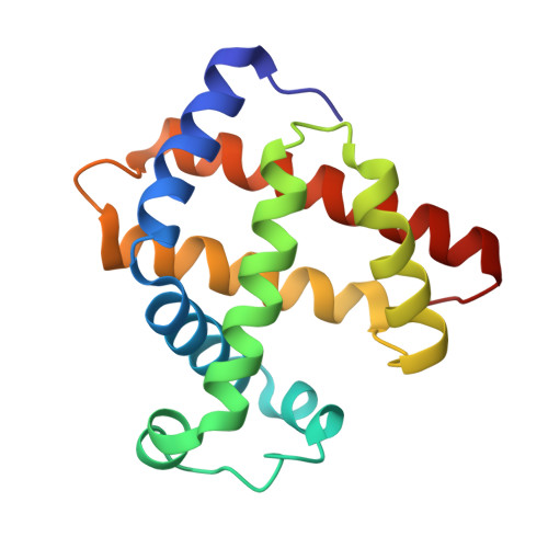
    Apo-myoglobin (apo-Mb) and apo-A71GMb were successfully reconstituted with FeIII(salophen) (1) (salophen = N,N'-bis(salicylidene)-1,2-phenilenediamine), Fe(III)(3,3'-Me2-salophen) (2), and FeIII(5,5'-t-Bu2-salophen) (3). The crystal structure of 2.apo-A71GMb shows the tight binding of the complex in the Mb cavity, while in wild-type apo-Mb it is highly disordered due to the steric repulsion with Ala71. Furthermore, the structure of 2.apo-A71GMb suggests a possible accommodation of a small substrate in the cavity. In fact, the cyanide association rate constant of 2.apo-A71GMb is 216-fold larger compared to that of 2.apo-Mb. These results provide us principles for the noncovalent fixation of synthetic metal cofactors at the desired positions in protein matrixes.


  • Organizational Affiliation
    • Research Center for Materials Science, Nagoya University, Nagoya 464-8602, Japan.

Macromolecule Content 

  • Total Structure Weight: 18.32 kDa 
  • Atom Count: 1,484 
  • Modeled Residue Count: 154 
  • Deposited Residue Count: 154 
  • Unique protein chains: 1

Macromolecules

Find similar proteins by:|  3D Structure
Entity ID: 1
MoleculeChains  Sequence LengthOrganismDetailsImage
MYOGLOBIN154Physeter macrocephalusMutation(s): 1 
EC: 1.11.1 (UniProt), 1.7 (UniProt)
UniProt
Find proteins for P02185 (Physeter macrocephalus)
Explore P02185 
Go to UniProtKB:  P02185
Entity Groups
Sequence Clusters30% Identity50% Identity70% Identity90% Identity95% Identity100% Identity
UniProt GroupP02185
Sequence Annotations
Expand
Reference Sequence

Small Molecules

Ligands 3 Unique
IDChains Name / Formula / InChI Key2D Diagram3D Interactions
CZM

Query on CZM


Download:Ideal Coordinates CCD File
I [auth A]'N,N'-BIS-(2-HYDROXY-3-METHYL-BENZYLIDENE)-BENZENE-1,2-DIAMINE'
C22 H20 N2 O2
DNVUYVBRNCWGAK-RNIAWFEPSA-N
PO4

Query on PO4


Download:Ideal Coordinates CCD File
C [auth A]
D [auth A]
E [auth A]
F [auth A]
G [auth A]
C [auth A],
D [auth A],
E [auth A],
F [auth A],
G [auth A],
H [auth A]
PHOSPHATE ION
O4 P
NBIIXXVUZAFLBC-UHFFFAOYSA-K
FE

Query on FE


Download:Ideal Coordinates CCD File
B [auth A]FE (III) ION
Fe
VTLYFUHAOXGGBS-UHFFFAOYSA-N

Experimental Data & Validation

Experimental Data

  • Method: X-RAY DIFFRACTION
  • Resolution: 1.60 Å
  • R-Value Free:  0.212 (Depositor) 
  • R-Value Work:  0.198 (Depositor) 
Space Group: P 21 21 21
Unit Cell:
Length ( Å )Angle ( ˚ )
a = 33.268α = 90
b = 57.6β = 90
c = 75.296γ = 90
Software Package:
Software NamePurpose
CNSrefinement
DENZOdata reduction
SCALEPACKdata scaling
CNSphasing

Structure Validation

View Full Validation Report


Entry History 

Deposition Data

Revision History  (Full details and data files)

  • Version 1.0: 2004-05-18
    Type: Initial release
  • Version 1.1: 2008-04-27
    Changes: Version format compliance
  • Version 1.2: 2011-07-13
    Changes: Version format compliance
  • Version 1.3: 2021-11-10
    Changes: Database references, Derived calculations
  • Version 1.4: 2023-10-25
    Changes: Data collection, Refinement description