1UEK | pdb_00001uek

Crystal structure of 4-(cytidine 5'-diphospho)-2C-methyl-D-erythritol kinase


Experimental Data Snapshot

  • Method: X-RAY DIFFRACTION
  • Resolution: 1.70 Å
  • R-Value Free: 
    0.218 (Depositor), 0.196 (DCC) 
  • R-Value Work: 
    0.182 (Depositor) 
  • R-Value Observed: 
    0.184 (Depositor) 

wwPDB Validation 3D Report Full Report

Validation slider image for 1UEK

This is version 1.3 of the entry. See complete history

Literature

Crystal Structure of 4-(Cytidine 5'-diphospho)-2-C-methyl-D-erythritol kinase, an Enzyme in the Non-mevalonate Pathway of Isoprenoid Synthesis.

Wada, T.Kuzuyama, T.Satoh, S.Kuramitsu, S.Yokoyama, S.Unzai, S.Tame, J.R.Park, S.Y.

(2003) J Biological Chem 278: 30022-30027

  • DOI: https://doi.org/10.1074/jbc.M304339200
  • Primary Citation Related Structures: 
    1UEK

  • PubMed Abstract: 

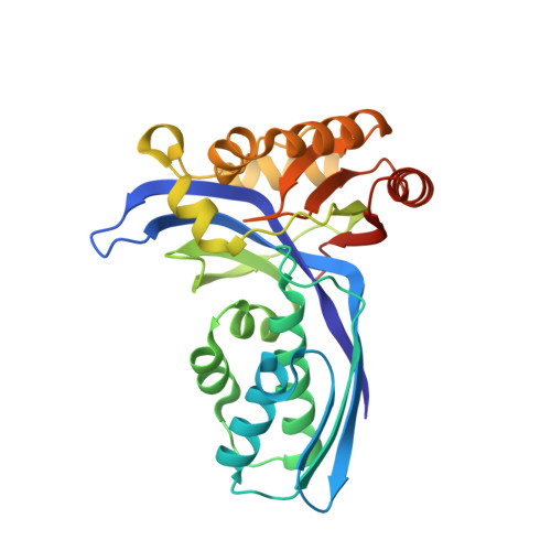
    The crystal structure of the enzyme 4-(cytidine 5'-diphospho)-2-C-methyl-D-erythritol (CDP-ME) kinase from the thermophilic bacterium Thermus thermophilus HB8 has been determined at 1.7-A resolution. This enzyme catalyzes phosphorylation of the 2-hydroxyl group of CDP-ME, the fourth step of the non-mevalonate pathway, which is essential for isoprenoid biosynthesis in several pathogenic microorganisms. Since this pathway is absent in humans, it is an important target for the development of novel antimicrobial compounds. The structure of the enzyme is similar to the structures of mevalonate kinase and homoserine kinase, members of the GHMP superfamily. Lys8 and Asp125 are active site residues in mevalonate kinase that also appear to play a catalytic role in CDP-ME kinase. Both the mevalonate and the non-mevalonate pathways therefore involve closely related kinases with similar mechanisms. Assaying the enzyme showed that CDP-ME kinase will phosphorylate CDP-ME but not 4-(uridine 5'-diphospho)-2-C-methyl-D-erythritol, indicating the substrate pyrimidine moiety is involved in important interactions with the enzyme.


  • Organizational Affiliation
    • Genomic Sciences Center, RIKEN Yokohama Institute, and Protein Design Laboratory, Yokohama City University, 1-7-29 Suehiro-cho, Tsurumi, Yokohama 230-0045, Japan.

Macromolecule Content 

  • Total Structure Weight: 29.29 kDa 
  • Atom Count: 2,230 
  • Modeled Residue Count: 268 
  • Deposited Residue Count: 275 
  • Unique protein chains: 1

Macromolecules

Find similar proteins by:|  3D Structure
Entity ID: 1
MoleculeChains  Sequence LengthOrganismDetailsImage
4-(cytidine 5'-diphospho)-2C-methyl-D-erythritol kinase275Thermus thermophilusMutation(s): 0 
Gene Names: ychB
EC: 2.7.1.148
UniProt
Find proteins for P83700 (Thermus thermophilus (strain ATCC 27634 / DSM 579 / HB8))
Explore P83700 
Go to UniProtKB:  P83700
Entity Groups
Sequence Clusters30% Identity50% Identity70% Identity90% Identity95% Identity100% Identity
UniProt GroupP83700
Sequence Annotations
Expand
Reference Sequence

Experimental Data & Validation

Experimental Data

  • Method: X-RAY DIFFRACTION
  • Resolution: 1.70 Å
  • R-Value Free:  0.218 (Depositor), 0.196 (DCC) 
  • R-Value Work:  0.182 (Depositor) 
  • R-Value Observed: 0.184 (Depositor) 
Space Group: P 21 21 21
Unit Cell:
Length ( Å )Angle ( ˚ )
a = 48.603α = 90
b = 66.21β = 90
c = 76.4γ = 90
Software Package:
Software NamePurpose
REFMACrefinement
HKL-2000data reduction
SCALEPACKdata scaling
SOLVEphasing

Structure Validation

View Full Validation Report


Entry History 

Deposition Data

Revision History  (Full details and data files)

  • Version 1.0: 2003-06-17
    Type: Initial release
  • Version 1.1: 2008-04-27
    Changes: Version format compliance
  • Version 1.2: 2011-07-13
    Changes: Version format compliance
  • Version 1.3: 2023-12-27
    Changes: Data collection, Database references