Crystal Structures and Structural Stabilities of the Disulfide Bond-Deficient Soybean Proglycinin Mutants C12G and C88S.
Adachi, M., Okuda, E., Kaneda, Y., Hashimoto, A., Shutov, A.D., Becker, C., Utsumi, S.(2003) J Agric Food Chem 51: 4633-4639
- PubMed: 14705889 Search on PubMed
- DOI: https://doi.org/10.1021/jf026065y
- Primary Citation Related Structures:
1UCX, 1UD1 - PubMed Abstract:
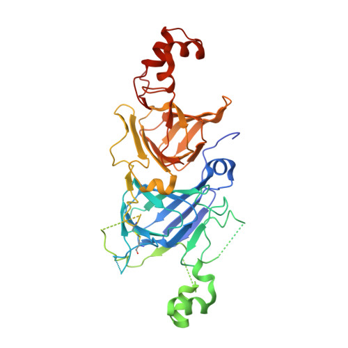
The constituent subunits of seed storage protein 11S globulin have two disulfide bonds that are common among 11S globulins from legume and nonlegume seeds. In the case of the A1aB1b subunit of soybean 11S globulin, glycinin, Cys12-Cys45 and Cys88-Cys298 are observed by X-ray crystallography. The significance of these two disulfide bonds for structural stability was investigated by mutagenesis of Cys12 to Gly and of Cys88 to Ser. The disulfide bond-deficient mutants C12G and C88S could form the correct conformations identical to that of the wild-type proglycinin except in the vicinities of the mutation sites C12 and C88 as shown by their crystal structures. Thermal stability monitored by differential scanning calorimetry of the mutants indicated that the contribution of these disulfide bonds to the thermal stability of proglycinin A1aB1b is low, although there is a small difference in the extent of the contribution between the two disulfide bonds (Cys12-Cys45 > Cys88-Cys298). The contribution of Cys88-Cys298 to the resistance of proglycinin A1aB1b to proteinase digestion is higher than that of Cys12-Cys45. Possible effects of structure on the different properties of C12G and C88S are discussed.
- Laboratory of Food Quality Design and Development, Graduate School of Agriculture, Kyoto University, Uji, Kyoto 611-0011, Japan.
Organizational Affiliation: