1UC7

Crystal structure of DsbDgamma


Experimental Data Snapshot

  • Method: X-RAY DIFFRACTION
  • Resolution: 1.90 Å
  • R-Value Free: 0.218 
  • R-Value Work: 0.185 
  • R-Value Observed: 0.191 

wwPDB Validation   3D Report Full Report


This is version 1.3 of the entry. See complete history


Literature

Crystal structure of DsbDgamma reveals the mechanism of redox potential shift and substrate specificity(1)

Kim, J.H.Kim, S.J.Jeong, D.G.Son, J.H.Ryu, S.E.

(2003) FEBS Lett 543: 164-169

  • DOI: https://doi.org/10.1016/s0014-5793(03)00434-4
  • Primary Citation of Related Structures:  
    1UC7

  • PubMed Abstract: 

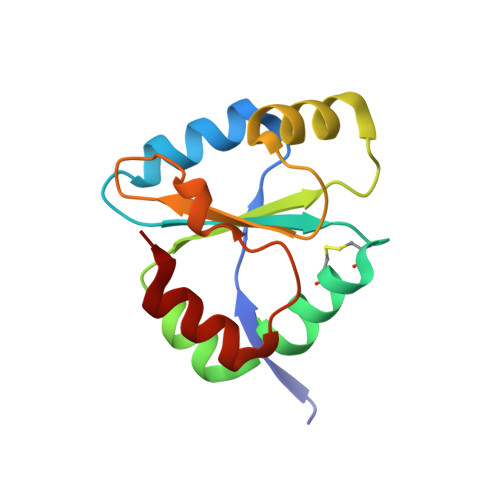
    The Escherichia coli transmembrane protein DsbD transfers electrons from the cytoplasm to the periplasm through a cascade of thiol-disulfide exchange reactions. In this process, the C-terminal periplasmic domain of DsbD (DsbDgamma) shuttles the reducing potential from the membrane domain (DsbDbeta) to the N-terminal periplasmic domain (DsbDalpha). The crystal structure of DsbDgamma determined at 1.9 A resolution reveals that the domain has a thioredoxin fold with an extended N-terminal stretch. In comparison to thioredoxin, the DsbDgamma structure exhibits the stabilized active site conformation and the extended active site alpha2 helix that explain the domain's substrate specificity and the redox potential shift, respectively. The hypothetical model of the DsbDgamma:DsbDalpha complex based on the DsbDgamma structure and previous structural studies indicates that the conserved hydrophobic residue in the C-X-X-C motif of DsbDgamma may be important in the specific recognition of DsbDalpha.


  • Organizational Affiliation

    Center for Cellular Switch Protein Structure, Korea Research Institute of Bioscience and Biotechnology, 52 Euh-eun-dong, Yusong-gu, 305-806, Daejon, South Korea.


Macromolecules
Find similar proteins by:  (by identity cutoff)  |  3D Structure
Entity ID: 1
MoleculeChains Sequence LengthOrganismDetailsImage
Thiol:disulfide interchange protein dsbD
A, B
125Escherichia coliMutation(s): 0 
EC: 1.8.1.8
UniProt
Find proteins for P58162 (Escherichia coli O157:H7)
Explore P58162 
Go to UniProtKB:  P58162
Entity Groups  
Sequence Clusters30% Identity50% Identity70% Identity90% Identity95% Identity100% Identity
UniProt GroupP58162
Sequence Annotations
Expand
  • Reference Sequence
Experimental Data & Validation

Experimental Data

  • Method: X-RAY DIFFRACTION
  • Resolution: 1.90 Å
  • R-Value Free: 0.218 
  • R-Value Work: 0.185 
  • R-Value Observed: 0.191 
  • Space Group: P 21 21 21
Unit Cell:
Length ( Å )Angle ( ˚ )
a = 30.33α = 90
b = 57.66β = 90
c = 126.6γ = 90
Software Package:
Software NamePurpose
MOSFLMdata reduction
SCALAdata scaling
SHARPphasing
CNSrefinement
CCP4data scaling

Structure Validation

View Full Validation Report


Entry History 

Deposition Data

Revision History  (Full details and data files)

  • Version 1.0: 2004-04-27
    Type: Initial release
  • Version 1.1: 2008-04-27
    Changes: Version format compliance
  • Version 1.2: 2011-07-13
    Changes: Version format compliance
  • Version 1.3: 2023-12-27
    Changes: Data collection, Database references