1UC5

Structure of diol dehydratase complexed with (R)-1,2-propanediol


Experimental Data Snapshot

  • Method: X-RAY DIFFRACTION
  • Resolution: 2.30 Å
  • R-Value Free: 0.255 
  • R-Value Work: 0.208 
  • R-Value Observed: 0.209 

wwPDB Validation   3D Report Full Report


Ligand Structure Quality Assessment 


This is version 2.1 of the entry. See complete history


Literature

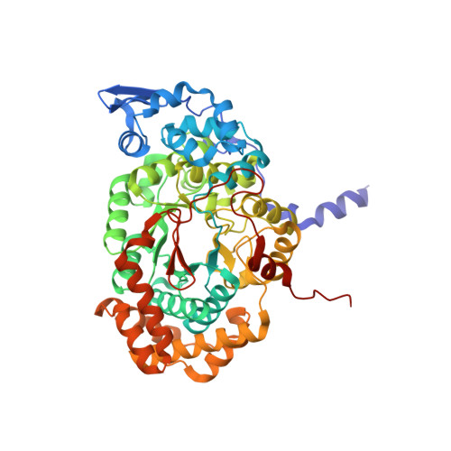
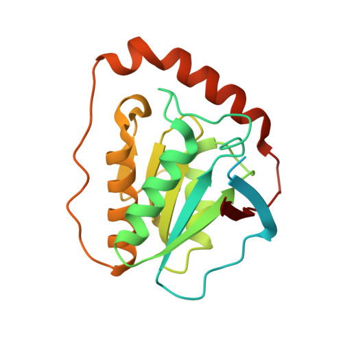
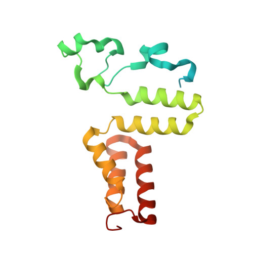
Structural rationalization for the lack of stereospecificity in coenzyme B12-dependent diol dehydratase

Shibata, N.Nakanishi, Y.Fukuoka, M.Yamanishi, M.Yasuoka, N.Toraya, T.

(2003) J Biol Chem 278: 22717-22725

  • DOI: https://doi.org/10.1074/jbc.M301513200
  • Primary Citation of Related Structures:  
    1UC4, 1UC5

  • PubMed Abstract: 

    Adenosylcobalamin-dependent diol dehydratase of Klebsiella oxytoca is apparently not stereospecific and catalyzes the conversion of both (R)- and (S)-1,2-propanediol to propionaldehyde. To explain this unusual property of the enzyme, we analyzed the crystal structures of diol dehydratase in complexes with cyanocobalamin and (R)- or (S)-1,2-propanediol. (R)- and (S)-isomers are bound in a symmetrical manner, although the hydrogen-bonding interactions between the substrate and the active-site residues are the same. From the position of the adenosyl radical in the modeled "distal" conformation, it is reasonable for the radical to abstract the pro-R and pro-S hydrogens from (R)- and (S)-isomers, respectively. The hydroxyl groups in the substrate radicals would migrates from C(2) to C(1) by a suprafacial shift, resulting in the stereochemical inversion at C(1). This causes 60 degrees clockwise and 70 degrees counterclockwise rotations of the C(1)-C(2) bond of the (R)- and (S)-isomers, respectively, if viewed from K+. A modeling study of 1,1-gem-diol intermediates indicated that new radical center C(2) becomes close to the methyl group of 5'-deoxyadenosine. Thus, the hydrogen back-abstraction (recombination) from 5'-deoxyadenosine by the product radical is structurally feasible. It was also predictable that the substitution of the migrating hydroxyl group by a hydrogen atom from 5'-deoxyadenosine takes place with the inversion of the configuration at C(2) of the substrate. Stereospecific dehydration of the 1,1-gem-diol intermediates can also be rationalized by assuming that Asp-alpha335 and Glu-alpha170 function as base catalysts in the dehydration of the (R)- and (S)-isomers, respectively. The structure-based mechanism and stereochemical courses of the reaction are proposed.


  • Organizational Affiliation

    Department of Life Science, Graduate School of Science, Himeji Institute of Technology, 3-2-1 Kouto, Kamigori, Ako-gun, Hyogo 678-1297, Japan.


Macromolecules
Find similar proteins by:  (by identity cutoff)  |  3D Structure
Entity ID: 1
MoleculeChains Sequence LengthOrganismDetailsImage
diol dehydrase alpha subunitA,
D [auth L]
554Klebsiella oxytocaMutation(s): 0 
EC: 4.2.1.28
UniProt
Find proteins for Q59470 (Klebsiella oxytoca)
Explore Q59470 
Go to UniProtKB:  Q59470
Entity Groups  
Sequence Clusters30% Identity50% Identity70% Identity90% Identity95% Identity100% Identity
UniProt GroupQ59470
Sequence Annotations
Expand
  • Reference Sequence
Find similar proteins by:  (by identity cutoff)  |  3D Structure
Entity ID: 2
MoleculeChains Sequence LengthOrganismDetailsImage
diol dehydrase beta subunit
B, E
224Klebsiella oxytocaMutation(s): 0 
EC: 4.2.1.28
UniProt
Find proteins for Q59471 (Klebsiella oxytoca)
Explore Q59471 
Go to UniProtKB:  Q59471
Entity Groups  
Sequence Clusters30% Identity50% Identity70% Identity90% Identity95% Identity100% Identity
UniProt GroupQ59471
Sequence Annotations
Expand
  • Reference Sequence
Find similar proteins by:  (by identity cutoff)  |  3D Structure
Entity ID: 3
MoleculeChains Sequence LengthOrganismDetailsImage
diol dehydrase gamma subunitC [auth G],
F [auth M]
173Klebsiella oxytocaMutation(s): 0 
EC: 4.2.1.28
UniProt
Find proteins for Q59472 (Klebsiella oxytoca)
Explore Q59472 
Go to UniProtKB:  Q59472
Entity Groups  
Sequence Clusters30% Identity50% Identity70% Identity90% Identity95% Identity100% Identity
UniProt GroupQ59472
Sequence Annotations
Expand
  • Reference Sequence
Small Molecules
Ligands 4 Unique
IDChains Name / Formula / InChI Key2D Diagram3D Interactions
CNC
Query on CNC

Download Ideal Coordinates CCD File 
K [auth B],
P [auth E]
CYANOCOBALAMIN
C63 H89 Co N14 O14 P
SYZBZQWSWIJYAR-UVKKECPRSA-M
PGR
Query on PGR

Download Ideal Coordinates CCD File 
J [auth A],
O [auth L]
R-1,2-PROPANEDIOL
C3 H8 O2
DNIAPMSPPWPWGF-GSVOUGTGSA-N
K
Query on K

Download Ideal Coordinates CCD File 
G [auth A],
L
POTASSIUM ION
K
NPYPAHLBTDXSSS-UHFFFAOYSA-N
NH4
Query on NH4

Download Ideal Coordinates CCD File 
H [auth A],
I [auth A],
M [auth L],
N [auth L]
AMMONIUM ION
H4 N
QGZKDVFQNNGYKY-UHFFFAOYSA-O
Experimental Data & Validation

Experimental Data

  • Method: X-RAY DIFFRACTION
  • Resolution: 2.30 Å
  • R-Value Free: 0.255 
  • R-Value Work: 0.208 
  • R-Value Observed: 0.209 
  • Space Group: P 21 21 21
Unit Cell:
Length ( Å )Angle ( ˚ )
a = 74.64α = 90
b = 122.27β = 90
c = 207.43γ = 90
Software Package:
Software NamePurpose
HKL-2000data collection
SCALEPACKdata scaling
CNSrefinement
HKL-2000data reduction
CNSphasing

Structure Validation

View Full Validation Report


Ligand Structure Quality Assessment 


Entry History 

Deposition Data

Revision History  (Full details and data files)

  • Version 1.0: 2003-07-22
    Type: Initial release
  • Version 1.1: 2008-04-27
    Changes: Version format compliance
  • Version 1.2: 2011-07-13
    Changes: Version format compliance
  • Version 2.0: 2021-08-18
    Changes: Data collection, Database references, Derived calculations, Non-polymer description, Structure summary
  • Version 2.1: 2023-10-25
    Changes: Data collection, Refinement description