1UBJ

Three-dimensional Structure of The Carbon Monoxide Complex of [NiFe]hydrogenase From Desulufovibrio vulgaris Miyazaki F


Experimental Data Snapshot

  • Method: X-RAY DIFFRACTION
  • Resolution: 1.35 Å
  • R-Value Free: 0.183 
  • R-Value Work: 0.121 
  • R-Value Observed: 0.124 

wwPDB Validation   3D Report Full Report


This is version 2.1 of the entry. See complete history


Literature

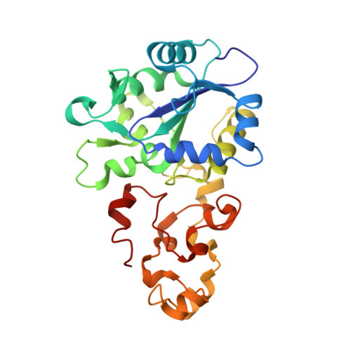
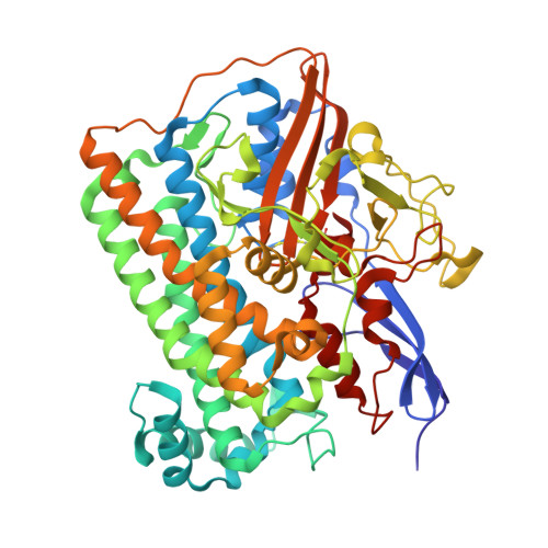
Structural Studies of the Carbon Monoxide Complex of [NiFe]hydrogenase from Desulfovibrio vulgaris Miyazaki F: Suggestion for the Initial Activation Site for Dihydrogen

Ogata, H.Mizoguchi, Y.Mizuno, N.Miki, K.Adachi, S.Yasuoka, N.Yagi, T.Yamauchi, O.Hirota, S.Higuchi, Y.

(2002) J Am Chem Soc 124: 11628-11635

  • DOI: https://doi.org/10.1021/ja012645k
  • Primary Citation of Related Structures:  
    1UBH, 1UBJ, 1UBK, 1UBL, 1UBM, 1UBO, 1UBR, 1UBT, 1UBU

  • PubMed Abstract: 

    The carbon monoxide complex of [NiFe]hydrogenase from Desulfovibrio vulgaris Miyazaki F has been characterized by X-ray crystallography and absorption and resonance Raman spectroscopy. Nine crystal structures of the [NiFe]hydrogenase in the CO-bound and CO-liberated forms were determined at 1.2-1.4 A resolution. The exogenously added CO was assigned to be bound to the Ni atom at the Ni-Fe active site. The CO was not replaced with H(2) in the dark at 100 K, but was liberated by illumination with a strong white light. The Ni-C distances and Ni-C-O angles were about 1.77 A and 160 degrees, respectively, except for one case (1.72 A and 135 degrees ), in which an additional electron density peak between the CO and Sgamma(Cys546) was recognized. Distinct changes were observed in the electron density distribution of the Ni and Sgamma(Cys546) atoms between the CO-bound and CO-liberated structures for all the crystals tested. The novel structural features found near the Ni and Sgamma(Cys546) atoms suggest that these two atoms at the Ni-Fe active site play a role during the initial H(2)-binding process. Anaerobic addition of CO to dithionite-reduced [NiFe]hydrogenase led to a new absorption band at about 470 nm ( approximately 3000 M(-1)cm(-1)). Resonance Raman spectra (excitation at 476.5 nm) of the CO complex revealed CO-isotope-sensitive bands at 375/393 and 430 cm(-1) (368 and 413 cm(-1) for (13)C(18)O). The frequencies and relative intensities of the CO-related Raman bands indicated that the exogenous CO is bound to the Ni atom with a bent Ni-C-O structure in solution, in agreement with the refined structure determined by X-ray crystallography.


  • Organizational Affiliation

    Division of Chemistry, Graduate School of Science, Kyoto University, Sakyo-ku, Kyoto 606-8502, Japan.


Macromolecules
Find similar proteins by:  (by identity cutoff)  |  3D Structure
Entity ID: 1
MoleculeChains Sequence LengthOrganismDetailsImage
Periplasmic [NiFe] hydrogenase small subunitA [auth S]267Nitratidesulfovibrio vulgaris str. 'Miyazaki FMutation(s): 0 
EC: 1.12.2.1
UniProt
Find proteins for P21853 (Desulfovibrio vulgaris (strain DSM 19637 / Miyazaki F))
Explore P21853 
Go to UniProtKB:  P21853
Entity Groups  
Sequence Clusters30% Identity50% Identity70% Identity90% Identity95% Identity100% Identity
UniProt GroupP21853
Sequence Annotations
Expand
  • Reference Sequence
Find similar proteins by:  (by identity cutoff)  |  3D Structure
Entity ID: 2
MoleculeChains Sequence LengthOrganismDetailsImage
Periplasmic [NiFe] hydrogenase large subunitB [auth L]534Nitratidesulfovibrio vulgaris str. 'Miyazaki FMutation(s): 0 
EC: 1.12.2.1
UniProt
Find proteins for P21852 (Desulfovibrio vulgaris (strain DSM 19637 / Miyazaki F))
Explore P21852 
Go to UniProtKB:  P21852
Entity Groups  
Sequence Clusters30% Identity50% Identity70% Identity90% Identity95% Identity100% Identity
UniProt GroupP21852
Sequence Annotations
Expand
  • Reference Sequence
Small Molecules
Ligands 5 Unique
IDChains Name / Formula / InChI Key2D Diagram3D Interactions
SF4
Query on SF4

Download Ideal Coordinates CCD File 
C [auth S],
D [auth S]
IRON/SULFUR CLUSTER
Fe4 S4
LJBDFODJNLIPKO-UHFFFAOYSA-N
F3S
Query on F3S

Download Ideal Coordinates CCD File 
E [auth S]FE3-S4 CLUSTER
Fe3 S4
FCXHZBQOKRZXKS-UHFFFAOYSA-N
FNE
Query on FNE

Download Ideal Coordinates CCD File 
G [auth L](MU-SULPHIDO)-BIS(MU-CYS,S)-[TRICARBONYLIRON-DI-(CYS,S)NICKEL(II)](FE-NI)
C3 Fe Ni O3 S
RSNJUOPWNIKDNA-UHFFFAOYSA-N
CMO
Query on CMO

Download Ideal Coordinates CCD File 
H [auth L]CARBON MONOXIDE
C O
UGFAIRIUMAVXCW-UHFFFAOYSA-N
MG
Query on MG

Download Ideal Coordinates CCD File 
F [auth L]MAGNESIUM ION
Mg
JLVVSXFLKOJNIY-UHFFFAOYSA-N
Experimental Data & Validation

Experimental Data

  • Method: X-RAY DIFFRACTION
  • Resolution: 1.35 Å
  • R-Value Free: 0.183 
  • R-Value Work: 0.121 
  • R-Value Observed: 0.124 
  • Space Group: P 21 21 21
Unit Cell:
Length ( Å )Angle ( ˚ )
a = 97.74α = 90
b = 125.86β = 90
c = 66.57γ = 90
Software Package:
Software NamePurpose
DENZOdata reduction
SCALAdata scaling
CNSrefinement
SHELXL-97refinement
CCP4data scaling
CNSphasing

Structure Validation

View Full Validation Report


Entry History 

Deposition Data

Revision History  (Full details and data files)

  • Version 1.0: 2003-04-29
    Type: Initial release
  • Version 1.1: 2008-04-27
    Changes: Version format compliance
  • Version 1.2: 2011-07-13
    Changes: Version format compliance
  • Version 2.0: 2019-08-28
    Changes: Atomic model, Data collection, Derived calculations
  • Version 2.1: 2023-10-25
    Changes: Data collection, Database references, Derived calculations, Refinement description