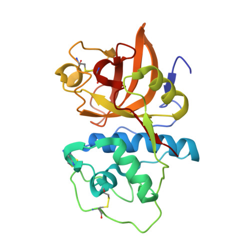
1U9V

Crystal Structure of the Cysteine Protease Human Cathepsin K in Complex with the Covalent Inhibitor NVP-ABE854


Experimental Data Snapshot

  • Method: X-RAY DIFFRACTION
  • Resolution: 2.20 Å
  • R-Value Free: 0.231 
  • R-Value Work: 0.170 
  • R-Value Observed: 0.170 

wwPDB Validation   3D Report Full Report


This is version 1.4 of the entry. See complete history


Literature

Novel purine nitrile derived inhibitors of the cysteine protease cathepsin K

Altmann, E.Cowan-Jacob, S.W.Missbach, M.

(2004) J Med Chem 47: 5833-5836

  • DOI: https://doi.org/10.1021/jm0493111
  • Primary Citation of Related Structures:  
    1U9V, 1U9W, 1U9X

  • PubMed Abstract: 

    Starting from the high-throughput screening hit 1a, novel cathepsin K inhibitors have been developed based on a purine scaffold. High-resolution X-ray structures of several derivatives have revealed the binding mode of these unique cysteine protease inhibitors.


  • Organizational Affiliation

    Novartis Institutes for BioMedical Research, CH-4002 Basel, Switzerland. eva.altmann@pharma.novartis.com


Macromolecules
Find similar proteins by:  (by identity cutoff)  |  3D Structure
Entity ID: 1
MoleculeChains Sequence LengthOrganismDetailsImage
Cathepsin K217Homo sapiensMutation(s): 0 
Gene Names: CTSKCTSOCTSO2
EC: 3.4.22.38
UniProt & NIH Common Fund Data Resources
Find proteins for P43235 (Homo sapiens)
Explore P43235 
Go to UniProtKB:  P43235
PHAROS:  P43235
Entity Groups  
Sequence Clusters30% Identity50% Identity70% Identity90% Identity95% Identity100% Identity
UniProt GroupP43235
Sequence Annotations
Expand
  • Reference Sequence
Small Molecules
Ligands 1 Unique
IDChains Name / Formula / InChI Key2D Diagram3D Interactions
IHE
Query on IHE

Download Ideal Coordinates CCD File 
B [auth A]6-(CYCLOHEXYLAMINO)-9-[2-(4-METHYLPIPERAZIN-1-YL)-ETHYL]-9H-PURINE-2-CARBONITRILE
C19 H28 N8
OGODDSLNRULSMM-UHFFFAOYSA-N
Binding Affinity Annotations 
IDSourceBinding Affinity
IHE PDBBind:  1U9V IC50: 6 (nM) from 1 assay(s)
BindingDB:  1U9V IC50: 6 (nM) from 1 assay(s)
Experimental Data & Validation

Experimental Data

  • Method: X-RAY DIFFRACTION
  • Resolution: 2.20 Å
  • R-Value Free: 0.231 
  • R-Value Work: 0.170 
  • R-Value Observed: 0.170 
  • Space Group: P 43 21 2
Unit Cell:
Length ( Å )Angle ( ˚ )
a = 63.2α = 90
b = 63.2β = 90
c = 113.8γ = 90
Software Package:
Software NamePurpose
XDSdata scaling
AUTOMARdata reduction
AMoREphasing
X-PLORrefinement
XDSdata reduction

Structure Validation

View Full Validation Report


Entry History 

Deposition Data

Revision History  (Full details and data files)

  • Version 1.0: 2005-08-11
    Type: Initial release
  • Version 1.1: 2008-04-30
    Changes: Version format compliance
  • Version 1.2: 2011-07-13
    Changes: Version format compliance
  • Version 1.3: 2017-10-11
    Changes: Refinement description
  • Version 1.4: 2023-10-25
    Changes: Data collection, Database references, Derived calculations, Refinement description