X-ray Crystal Structure of the Hypothetical Phosphotyrosine Phosphatase MDP-1 of the Haloacid Dehalogenase Superfamily
Peisach, E., Selengut, J.D., Dunaway-Mariano, D., Allen, K.N.(2004) Biochemistry 43: 12770-12779
- PubMed: 15461449 Search on PubMed
- DOI: https://doi.org/10.1021/bi0490688
- Primary Citation Related Structures:
1U7O, 1U7P - PubMed Abstract:
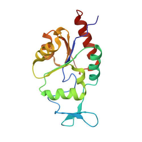
The haloacid dehalogenase (HAD) superfamily is comprised of structurally homologous enzymes that share several conserved sequence motifs (loops I-IV) in their active site. The majority of HAD members are phosphohydrolases and may be divided into three subclasses depending on domain organization. In classes I and II, a mobile "cap" domain reorients upon substrate binding, closing the active site to bulk solvent. Members of the third class lack this additional domain. Herein, we report the 1.9 A X-ray crystal structures of a member of the third subclass, magnesium-dependent phosphatase-1 (MDP-1) both in its unliganded form and with the product analogue, tungstate, bound to the active site. The secondary structure of MDP-1 is similar to that of the "core" domain of other type I and type II HAD members with the addition of a small, 28-amino acid insert that does not close down to exclude bulk solvent in the presence of ligand. In addition, the monomeric oligomeric state of MDP-1 does not allow the participation of a second subunit in the formation and solvent protection of the active site. The binding sites for the phosphate portion of the substrate and Mg(II) cofactor are also similar to those of other HAD members, with all previously observed contacts conserved. Unlike other subclass III HAD members, MDP-1 appears to be equally able to dephosphorylate phosphotyrosine and closed-ring phosphosugars. Modeling of possible substrates in the active site of MDP-1 reveals very few potential interactions with the substrate leaving group. The mapping of conserved residues in sequences of MDP-1 from different eukaryotic organisms reveals that they colocalize to a large region on the surface of the protein outside the active site. This observation combined with the modeling studies suggests that the target of MDP-1 is most likely a phosphotyrosine in an unknown protein rather than a small sugar-based substrate.
- Department of Physiology and Biophysics, Boston University School of Medicine, Boston, Massachusetts 02118-2394, USA.
Organizational Affiliation: