1U2G

transhydrogenase (dI.ADPr)2(dIII.NADPH)1 asymmetric complex


Experimental Data Snapshot

  • Method: X-RAY DIFFRACTION
  • Resolution: 2.20 Å
  • R-Value Free: 0.254 
  • R-Value Work: 0.222 

wwPDB Validation   3D Report Full Report


This is version 1.3 of the entry. See complete history


Literature

Active-site conformational changes associated with hydride transfer in proton-translocating transhydrogenase.

Mather, O.C.Singh, A.van Boxel, G.I.White, S.A.Jackson, J.B.

(2004) Biochemistry 43: 10952-10964

  • DOI: https://doi.org/10.1021/bi0497594
  • Primary Citation of Related Structures:  
    1U28, 1U2D, 1U2G, 1U31

  • PubMed Abstract: 

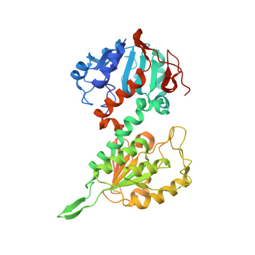
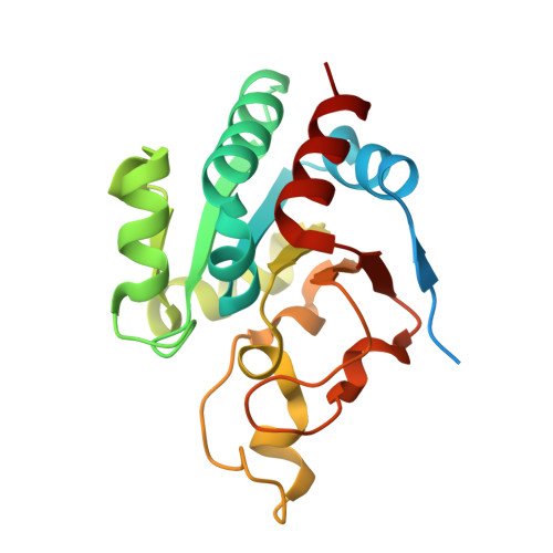
    Transhydrogenase couples the redox (hydride-transfer) reaction between NAD(H) and NADP(H) to proton translocation across a membrane. The redox reaction is catalyzed at the interface between two components (dI and dIII) which protrude from the membrane. A complex formed from recombinant dI and dIII (the dI(2)dIII(1) complex) from Rhodospirillum rubrum transhydrogenase catalyzes fast single-turnover hydride transfer between bound nucleotides. In this report we describe three new crystal structures of the dI(2)dIII(1) complex in different nucleotide-bound forms. The structures reveal an asymmetry in nucleotide binding that complements results from solution studies and supports the notion that intact transhydrogenase functions by an alternating site mechanism. In one structure, the redox site is occupied by NADH (on dI) and NADPH (on dIII). The dihydronicotinamide rings take up positions which may approximate to the ground state for hydride transfer: the redox-active C4(N) atoms are separated by only 3.6 A, and the perceived reaction stereochemistry matches that observed experimentally. The NADH conformation is different in the two dI polypeptides of this form of the dI(2)dIII(1) complex. Comparisons between a number of X-ray structures show that a conformational change in the NADH is driven by relative movement of the two domains which comprise dI. It is suggested that an equivalent conformational change in the intact enzyme is important in gating the hydride-transfer reaction. The observed nucleotide conformational change in the dI(2)dIII(1) complex is accompanied by rearrangements in the orientation of local amino acid side chains which may be responsible for sealing the site from the solvent and polarizing hydride transfer.


  • Organizational Affiliation

    School of Biosciences, University of Birmingham, Edgbaston, Birmingham B15 2TT, UK.


Macromolecules
Find similar proteins by:  (by identity cutoff)  |  3D Structure
Entity ID: 1
MoleculeChains Sequence LengthOrganismDetailsImage
NAD(P) transhydrogenase subunit alpha part 1
A, B
384Rhodospirillum rubrumMutation(s): 0 
Gene Names: pntAAnntA1
EC: 1.6.1.2
UniProt
Find proteins for Q2RSB2 (Rhodospirillum rubrum (strain ATCC 11170 / ATH 1.1.1 / DSM 467 / LMG 4362 / NCIMB 8255 / S1))
Explore Q2RSB2 
Go to UniProtKB:  Q2RSB2
Entity Groups  
Sequence Clusters30% Identity50% Identity70% Identity90% Identity95% Identity100% Identity
UniProt GroupQ2RSB2
Sequence Annotations
Expand
  • Reference Sequence
Find similar proteins by:  (by identity cutoff)  |  3D Structure
Entity ID: 2
MoleculeChains Sequence LengthOrganismDetailsImage
NAD(P) transhydrogenase subunit beta203Rhodospirillum rubrumMutation(s): 0 
Gene Names: pntBnntB
EC: 1.6.1.2
UniProt
Find proteins for Q2RSB4 (Rhodospirillum rubrum (strain ATCC 11170 / ATH 1.1.1 / DSM 467 / LMG 4362 / NCIMB 8255 / S1))
Explore Q2RSB4 
Go to UniProtKB:  Q2RSB4
Entity Groups  
Sequence Clusters30% Identity50% Identity70% Identity90% Identity95% Identity100% Identity
UniProt GroupQ2RSB4
Sequence Annotations
Expand
  • Reference Sequence
Small Molecules
Ligands 3 Unique
IDChains Name / Formula / InChI Key2D Diagram3D Interactions
NDP
Query on NDP

Download Ideal Coordinates CCD File 
G [auth C]NADPH DIHYDRO-NICOTINAMIDE-ADENINE-DINUCLEOTIDE PHOSPHATE
C21 H30 N7 O17 P3
ACFIXJIJDZMPPO-NNYOXOHSSA-N
APR
Query on APR

Download Ideal Coordinates CCD File 
D [auth A],
E [auth B]
ADENOSINE-5-DIPHOSPHORIBOSE
C15 H23 N5 O14 P2
SRNWOUGRCWSEMX-KEOHHSTQSA-N
GOL
Query on GOL

Download Ideal Coordinates CCD File 
F [auth B]GLYCEROL
C3 H8 O3
PEDCQBHIVMGVHV-UHFFFAOYSA-N
Experimental Data & Validation

Experimental Data

  • Method: X-RAY DIFFRACTION
  • Resolution: 2.20 Å
  • R-Value Free: 0.254 
  • R-Value Work: 0.222 
  • Space Group: P 21 21 21
Unit Cell:
Length ( Å )Angle ( ˚ )
a = 72.356α = 90
b = 73.766β = 90
c = 205.026γ = 90
Software Package:
Software NamePurpose
MOSFLMdata reduction
SCALAdata scaling
PHASESphasing
REFMACrefinement
CCP4data scaling

Structure Validation

View Full Validation Report


Entry History 

Deposition Data

Revision History  (Full details and data files)

  • Version 1.0: 2005-01-25
    Type: Initial release
  • Version 1.1: 2008-04-30
    Changes: Version format compliance
  • Version 1.2: 2011-07-13
    Changes: Non-polymer description, Version format compliance
  • Version 1.3: 2023-08-23
    Changes: Data collection, Database references, Derived calculations, Refinement description