1U2C

Crystal Structure of a-dystroglycan


Experimental Data Snapshot

  • Method: X-RAY DIFFRACTION
  • Resolution: 2.30 Å
  • R-Value Free: 0.262 
  • R-Value Work: 0.208 
  • R-Value Observed: 0.208 

wwPDB Validation   3D Report Full Report


This is version 1.2 of the entry. See complete history


Literature

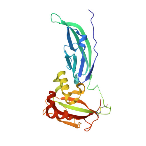
The Structure of the N-terminal Region of Murine Skeletal Muscle {alpha}-Dystroglycan Discloses a Modular Architecture

Bozic, D.Sciandra, F.Lamba, D.Brancaccio, A.

(2004) J Biol Chem 279: 44812-44816

  • DOI: https://doi.org/10.1074/jbc.C400353200
  • Primary Citation of Related Structures:  
    1U2C

  • PubMed Abstract: 

    Dystroglycan (DG) is a cell surface receptor consisting of two subunits: alpha-dystroglycan, extracellular and highly glycosylated, and beta-dystroglycan, spanning the cell membrane. It is a pivotal member of the dystrophin-glycoprotein complex and is involved in a wide variety of important cellular processes such as the stabilization of the muscle fiber sarcolemma or the clustering of acetylcholine receptors. We report the 2.3-A resolution crystal structure of the murine skeletal muscle N-terminal alpha-DG region, which confirms the presence of two autonomous domains; the first finally identified as an Ig-like and the second resembling ribosomal RNA-binding proteins. Solid-phase laminin binding assays show the occurrence of protein-protein type of interactions involving the Ig-like domain of alpha-DG.


  • Organizational Affiliation

    Biochemisches Institut der Universität Zürich, Zürich 8044, Switzerland. damir.bozic@mat.ethz.ch


Macromolecules
Find similar proteins by:  (by identity cutoff)  |  3D Structure
Entity ID: 1
MoleculeChains Sequence LengthOrganismDetailsImage
Dystroglycan246Mus musculusMutation(s): 0 
UniProt
Find proteins for Q62165 (Mus musculus)
Explore Q62165 
Go to UniProtKB:  Q62165
Entity Groups  
Sequence Clusters30% Identity50% Identity70% Identity90% Identity95% Identity100% Identity
UniProt GroupQ62165
Sequence Annotations
Expand
  • Reference Sequence
Experimental Data & Validation

Experimental Data

  • Method: X-RAY DIFFRACTION
  • Resolution: 2.30 Å
  • R-Value Free: 0.262 
  • R-Value Work: 0.208 
  • R-Value Observed: 0.208 
  • Space Group: H 3
Unit Cell:
Length ( Å )Angle ( ˚ )
a = 71.445α = 90
b = 71.445β = 90
c = 144.233γ = 120
Software Package:
Software NamePurpose
CNSrefinement
DENZOdata reduction
SCALEPACKdata scaling
CNSphasing

Structure Validation

View Full Validation Report


Entry History 

Deposition Data

Revision History  (Full details and data files)

  • Version 1.0: 2004-09-07
    Type: Initial release
  • Version 1.1: 2008-04-30
    Changes: Version format compliance
  • Version 1.2: 2011-07-13
    Changes: Version format compliance