1U0E

Crystal structure of mouse phosphoglucose isomerase


Experimental Data Snapshot

  • Method: X-RAY DIFFRACTION
  • Resolution: 1.60 Å
  • R-Value Free: 0.246 
  • R-Value Work: 0.211 
  • R-Value Observed: 0.213 

wwPDB Validation   3D Report Full Report


This is version 1.3 of the entry. See complete history


Literature

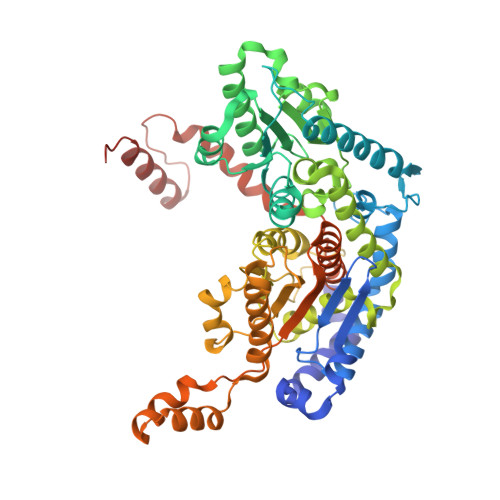
The crystal structure of mouse phosphoglucose isomerase at 1.6A resolution and its complex with glucose 6-phosphate reveals the catalytic mechanism of sugar ring opening.

Graham Solomons, J.T.Zimmerly, E.M.Burns, S.Krishnamurthy, N.Swan, M.K.Krings, S.Muirhead, H.Chirgwin, J.Davies, C.

(2004) J Mol Biol 342: 847-860

  • DOI: https://doi.org/10.1016/j.jmb.2004.07.085
  • Primary Citation of Related Structures:  
    1U0E, 1U0F, 1U0G

  • PubMed Abstract: 

    Phosphoglucose isomerase (PGI) is an enzyme of glycolysis that interconverts glucose 6-phosphate (G6P) and fructose 6-phosphate (F6P) but, outside the cell, is a multifunctional cytokine. High-resolution crystal structures of the enzyme from mouse have been determined in native form and in complex with the inhibitor erythrose 4-phosphate, and with the substrate glucose 6-phosphate. In the substrate-bound structure, the glucose sugar is observed in both straight-chain and ring forms. This structure supports a specific role for Lys518 in enzyme-catalyzed ring opening and we present a "push-pull" mechanism in which His388 breaks the O5-C1 bond by donating a proton to the ring oxygen atom and, simultaneously, Lys518 abstracts a proton from the C1 hydroxyl group. The reverse occurs in ring closure. The transition from ring form to straight-chain substrate is achieved through rotation of the C3-C4 bond, which brings the C1-C2 region into close proximity to Glu357, the base catalyst for the isomerization step. The structure with G6P also explains the specificity of PGI for glucose 6-phosphate over mannose 6-isomerase (M6P). To isomerize M6P to F6P requires a rotation of its C2-C3 bond but in PGI this is sterically blocked by Gln511.


  • Organizational Affiliation

    Department of Biochemistry and Molecular Biology, Medical University of South Carolina, Charleston, SC 29425, USA.


Macromolecules
Find similar proteins by:  (by identity cutoff)  |  3D Structure
Entity ID: 1
MoleculeChains Sequence LengthOrganismDetailsImage
Glucose-6-phosphate isomerase
A, B
564Mus musculusMutation(s): 0 
Gene Names: Gpi
EC: 5.3.1.9
UniProt
Find proteins for P06745 (Mus musculus)
Explore P06745 
Go to UniProtKB:  P06745
Entity Groups  
Sequence Clusters30% Identity50% Identity70% Identity90% Identity95% Identity100% Identity
UniProt GroupP06745
Sequence Annotations
Expand
  • Reference Sequence
Small Molecules
Ligands 3 Unique
IDChains Name / Formula / InChI Key2D Diagram3D Interactions
SO4
Query on SO4

Download Ideal Coordinates CCD File 
C [auth A]
D [auth A]
E [auth A]
F [auth A]
Q [auth B]
C [auth A],
D [auth A],
E [auth A],
F [auth A],
Q [auth B],
R [auth B],
S [auth B],
T [auth B],
U [auth B],
V [auth B]
SULFATE ION
O4 S
QAOWNCQODCNURD-UHFFFAOYSA-L
GOL
Query on GOL

Download Ideal Coordinates CCD File 
L [auth A]
M [auth A]
N [auth A]
O [auth A]
P [auth A]
L [auth A],
M [auth A],
N [auth A],
O [auth A],
P [auth A],
W [auth B],
X [auth B]
GLYCEROL
C3 H8 O3
PEDCQBHIVMGVHV-UHFFFAOYSA-N
BME
Query on BME

Download Ideal Coordinates CCD File 
G [auth A],
H [auth A],
I [auth A],
J [auth A],
K [auth A]
BETA-MERCAPTOETHANOL
C2 H6 O S
DGVVWUTYPXICAM-UHFFFAOYSA-N
Experimental Data & Validation

Experimental Data

  • Method: X-RAY DIFFRACTION
  • Resolution: 1.60 Å
  • R-Value Free: 0.246 
  • R-Value Work: 0.211 
  • R-Value Observed: 0.213 
  • Space Group: P 1 21 1
Unit Cell:
Length ( Å )Angle ( ˚ )
a = 69.2α = 90
b = 116.1β = 101.3
c = 73.1γ = 90
Software Package:
Software NamePurpose
REFMACrefinement
CrystalCleardata reduction
d*TREKdata scaling
MOLREPphasing

Structure Validation

View Full Validation Report


Entry History 

Deposition Data

Revision History  (Full details and data files)

  • Version 1.0: 2004-11-02
    Type: Initial release
  • Version 1.1: 2008-04-30
    Changes: Version format compliance
  • Version 1.2: 2011-07-13
    Changes: Non-polymer description, Version format compliance
  • Version 1.3: 2023-08-23
    Changes: Data collection, Database references, Derived calculations, Refinement description