1TZO

Crystal Structure of the Anthrax Toxin Protective Antigen Heptameric Prepore


Experimental Data Snapshot

  • Method: X-RAY DIFFRACTION
  • Resolution: 3.60 Å
  • R-Value Free: 0.317 
  • R-Value Work: 0.314 
  • R-Value Observed: 0.315 

wwPDB Validation   3D Report Full Report


This is version 1.4 of the entry. See complete history


Literature

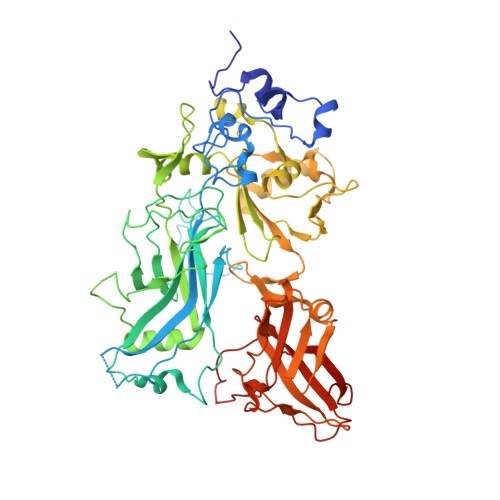
Structure of heptameric protective antigen bound to an anthrax toxin receptor: A role for receptor in pH-dependent pore formation

Lacy, D.B.Wigelsworth, D.J.Melnyk, R.A.Harrison, S.C.Collier, R.J.

(2004) Proc Natl Acad Sci U S A 101: 13147-13151

  • DOI: https://doi.org/10.1073/pnas.0405405101
  • Primary Citation of Related Structures:  
    1TZN, 1TZO

  • PubMed Abstract: 

    After binding to cellular receptors and proteolytic activation, the protective antigen component of anthrax toxin forms a heptameric prepore. The prepore later undergoes pH-dependent conversion to a pore, mediating translocation of the edema and lethal factors to the cytosol. We describe structures of the prepore (3.6 A) and a prepore:receptor complex (4.3 A) that reveal the location of pore-forming loops and an unexpected interaction of the receptor with the pore-forming domain. Lower pH is required for prepore-to-pore conversion in the presence of the receptor, indicating that this interaction regulates pH-dependent pore formation. We present an example of a receptor negatively regulating pH-dependent membrane insertion.


  • Organizational Affiliation

    Department of Microbiology and Molecular Genetics, Harvard Medical School, 200 Longwood Avenue, Boston, MA 02115, USA.


Macromolecules
Find similar proteins by:  (by identity cutoff)  |  3D Structure
Entity ID: 1
MoleculeChains Sequence LengthOrganismDetailsImage
Protective antigen
A,
B,
C,
D,
E,
F,
G,
H,
I,
J,
K,
L,
M,
N [auth O]
562Bacillus anthracis str. A2012Mutation(s): 0 
Gene Names: pagA
UniProt
Find proteins for P13423 (Bacillus anthracis)
Explore P13423 
Go to UniProtKB:  P13423
Entity Groups  
Sequence Clusters30% Identity50% Identity70% Identity90% Identity95% Identity100% Identity
UniProt GroupP13423
Sequence Annotations
Expand
  • Reference Sequence
Small Molecules
Ligands 1 Unique
IDChains Name / Formula / InChI Key2D Diagram3D Interactions
CA
Query on CA

Download Ideal Coordinates CCD File 
AA [auth G]
BA [auth G]
CA [auth H]
DA [auth H]
EA [auth I]
AA [auth G],
BA [auth G],
CA [auth H],
DA [auth H],
EA [auth I],
FA [auth I],
GA [auth J],
HA [auth J],
IA [auth K],
JA [auth K],
KA [auth L],
LA [auth L],
MA [auth M],
NA [auth M],
O [auth A],
OA [auth O],
P [auth A],
PA [auth O],
Q [auth B],
R [auth B],
S [auth C],
T [auth C],
U [auth D],
V [auth D],
W [auth E],
X [auth E],
Y [auth F],
Z [auth F]
CALCIUM ION
Ca
BHPQYMZQTOCNFJ-UHFFFAOYSA-N
Experimental Data & Validation

Experimental Data

  • Method: X-RAY DIFFRACTION
  • Resolution: 3.60 Å
  • R-Value Free: 0.317 
  • R-Value Work: 0.314 
  • R-Value Observed: 0.315 
  • Space Group: P 1
Unit Cell:
Length ( Å )Angle ( ˚ )
a = 149.608α = 77.56
b = 167.028β = 75.74
c = 168.2γ = 76.01
Software Package:
Software NamePurpose
DENZOdata reduction
SCALEPACKdata scaling
MOLREPphasing
CNSrefinement

Structure Validation

View Full Validation Report


Entry History 

Deposition Data

Revision History  (Full details and data files)

  • Version 1.0: 2004-08-17
    Type: Initial release
  • Version 1.1: 2008-04-30
    Changes: Version format compliance
  • Version 1.2: 2011-07-13
    Changes: Source and taxonomy, Version format compliance
  • Version 1.3: 2024-02-14
    Changes: Data collection, Database references, Derived calculations
  • Version 1.4: 2024-04-03
    Changes: Refinement description