1TXT

Staphylococcus aureus 3-hydroxy-3-methylglutaryl-CoA synthase


Experimental Data Snapshot

  • Method: X-RAY DIFFRACTION
  • Resolution: 2.50 Å
  • R-Value Free: 0.255 
  • R-Value Work: 0.187 
  • R-Value Observed: 0.191 

wwPDB Validation   3D Report Full Report


Ligand Structure Quality Assessment 


This is version 1.2 of the entry. See complete history


Literature

Staphylococcus aureus 3-hydroxy-3-methylglutaryl-CoA synthase: crystal structure and mechanism

Campobasso, N.Patel, M.Wilding, I.E.Kallender, H.Rosenberg, M.Gwynn, M.

(2004) J Biol Chem 279: 44883-44888

  • DOI: https://doi.org/10.1074/jbc.M407882200
  • Primary Citation of Related Structures:  
    1TVZ, 1TXT

  • PubMed Abstract: 

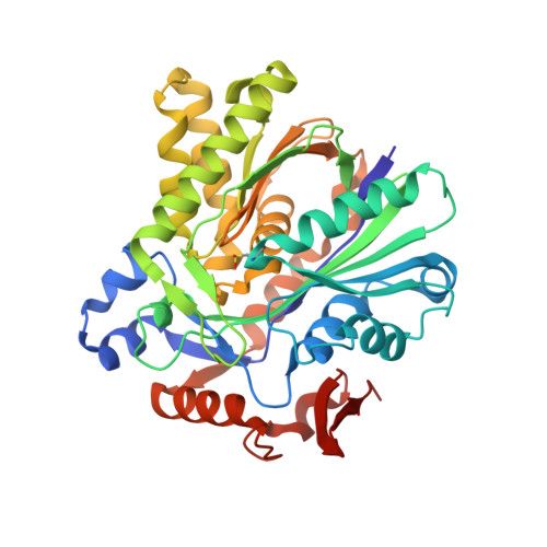
    3-Hydroxy-3-methylglutaryl coenzyme A (HMG-CoA) synthase, a member of the family of acyl-condensing enzymes, catalyzes the first committed step in the mevalonate pathway and is a potential target for novel antibiotics and cholesterol-lowering agents. The Staphylococcus aureus mvaS gene product (43.2 kDa) was overexpressed in Escherichia coli, purified to homogeneity, and shown biochemically to be an HMG-CoA synthase. The crystal structure of the full-length enzyme was determined at 2.0-A resolution, representing the first structure of an HMG-CoA synthase from any organism. HMG-CoA synthase forms a homodimer. The monomer, however, contains an important core structure of two similar alpha/beta motifs, a fold that is completely conserved among acyl-condensing enzymes. This common fold provides a scaffold for a catalytic triad made up of Cys, His, and Asn required by these enzymes. In addition, a crystal structure of HMG-CoA synthase with acetoacetyl-CoA was determined at 2.5-A resolution. Together, these structures provide the structural basis for an understanding of the mechanism of HMG-CoA synthase.


  • Organizational Affiliation

    GlaxoSmithKline Pharmaceuticals, Computational, Analytical, and Structural Sciences, King of Prussia, Pennsylvania 19406, USA. Nino.2.Campobasso@gsk.com


Macromolecules
Find similar proteins by:  (by identity cutoff)  |  3D Structure
Entity ID: 1
MoleculeChains Sequence LengthOrganismDetailsImage
3-hydroxy-3-methylglutaryl-CoA synthase
A, B, C, D
388Staphylococcus aureusMutation(s): 1 
Gene Names: mvaS
EC: 4.1.3.5
UniProt
Find proteins for Q9FD87 (Staphylococcus aureus)
Explore Q9FD87 
Go to UniProtKB:  Q9FD87
Entity Groups  
Sequence Clusters30% Identity50% Identity70% Identity90% Identity95% Identity100% Identity
UniProt GroupQ9FD87
Sequence Annotations
Expand
  • Reference Sequence
Experimental Data & Validation

Experimental Data

  • Method: X-RAY DIFFRACTION
  • Resolution: 2.50 Å
  • R-Value Free: 0.255 
  • R-Value Work: 0.187 
  • R-Value Observed: 0.191 
  • Space Group: P 1 21 1
Unit Cell:
Length ( Å )Angle ( ˚ )
a = 119.947α = 90
b = 64.482β = 97.17
c = 121.91γ = 90
Software Package:
Software NamePurpose
HKL-2000data collection
SCALEPACKdata scaling
AMoREphasing
REFMACrefinement
HKL-2000data reduction

Structure Validation

View Full Validation Report


Ligand Structure Quality Assessment 


Entry History 

Deposition Data

Revision History  (Full details and data files)

  • Version 1.0: 2004-08-31
    Type: Initial release
  • Version 1.1: 2008-04-30
    Changes: Version format compliance
  • Version 1.2: 2011-07-13
    Changes: Version format compliance