1TXK | pdb_00001txk

Crystal structure of Escherichia coli OpgG


Experimental Data Snapshot

  • Method: X-RAY DIFFRACTION
  • Resolution: 2.50 Å
  • R-Value Free: 
    0.229 (Depositor), 0.239 (DCC) 
  • R-Value Work: 
    0.192 (Depositor), 0.206 (DCC) 
  • R-Value Observed: 
    0.192 (Depositor) 

wwPDB Validation 3D Report Full Report

Validation slider image for 1TXK

This is version 1.3 of the entry. See complete history

Literature

Structural Analysis of Escherichia coli OpgG, a Protein Required for the Biosynthesis of Osmoregulated Periplasmic Glucans.

Hanoulle, X.Rollet, E.Clantin, B.Landrieu, I.Odberg-Ferragut, C.Lippens, G.Bohin, J.P.Villeret, V.

(2004) J Mol Biology 342: 195-205

  • DOI: https://doi.org/10.1016/j.jmb.2004.07.004
  • Primary Citation Related Structures: 
    1TXK

  • PubMed Abstract: 

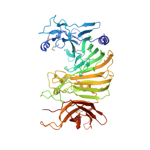
    Osmoregulated periplasmic glucans (OPGs) G protein (OpgG) is required for OPGs biosynthesis. OPGs from Escherichia coli are branched glucans, with a backbone of beta-1,2 glucose units and with branches attached by beta-1,6 linkages. In Proteobacteria, OPGs are involved in osmoprotection, biofilm formation, virulence and resistance to antibiotics. Despite their important biological implications, enzymes synthesizing OPGs are poorly characterized. Here, we report the 2.5 A crystal structure of OpgG from E.coli. The structure was solved using a selenemethionine derivative of OpgG and the multiple anomalous diffraction method (MAD). The protein is composed of two beta-sandwich domains connected by one turn of 3(10) helix. The N-terminal domain (residues 22-388) displays a 25-stranded beta-sandwich fold found in several carbohydrate-related proteins. It exhibits a large cleft comprising many aromatic and acidic residues. This putative binding site shares some similarities with enzymes such as galactose mutarotase and glucodextranase, suggesting a potential catalytic role for this domain in OPG synthesis. On the other hand, the C-terminal domain (residues 401-512) has a seven-stranded immunoglobulin-like beta-sandwich fold, found in many proteins where it is mainly implicated in interactions with other molecules. The structural data suggest that OpgG is an OPG branching enzyme in which the catalytic activity is located in the large N-terminal domain and controlled via the smaller C-terminal domain.


  • Organizational Affiliation
    • UMR 8525 CNRS, Institut de Biologie de Lille, Université de Lille II, 1 rue du Professeur Calmette, BP447, 59021, France. xavier.hanoulle@ibl.fr

Macromolecule Content 

  • Total Structure Weight: 114.2 kDa 
  • Atom Count: 8,291 
  • Modeled Residue Count: 977 
  • Deposited Residue Count: 996 
  • Unique protein chains: 1

Macromolecules

Find similar proteins by:|  3D Structure
Entity ID: 1
MoleculeChains  Sequence LengthOrganismDetailsImage
Glucans biosynthesis protein G
A, B
498Escherichia coliMutation(s): 10 
Gene Names: opgG
Membrane Entity: Yes 
UniProt
Find proteins for P33136 (Escherichia coli (strain K12))
Explore P33136 
Go to UniProtKB:  P33136
Entity Groups
Sequence Clusters30% Identity50% Identity70% Identity90% Identity95% Identity100% Identity
UniProt GroupP33136
Sequence Annotations
Expand
Reference Sequence

Small Molecules

Ligands 1 Unique
IDChains Name / Formula / InChI Key2D Diagram3D Interactions
NA

Query on NA


Download:Ideal Coordinates CCD File
C [auth A]SODIUM ION
Na
FKNQFGJONOIPTF-UHFFFAOYSA-N
Modified Residues  1 Unique
IDChains TypeFormula2D DiagramParent
MSE
Query on MSE
A, B
L-PEPTIDE LINKINGC5 H11 N O2 SeMET

Experimental Data & Validation

Experimental Data

  • Method: X-RAY DIFFRACTION
  • Resolution: 2.50 Å
  • R-Value Free:  0.229 (Depositor), 0.239 (DCC) 
  • R-Value Work:  0.192 (Depositor), 0.206 (DCC) 
  • R-Value Observed: 0.192 (Depositor) 
Space Group: P 21 21 21
Unit Cell:
Length ( Å )Angle ( ˚ )
a = 63.86α = 90
b = 88.12β = 90
c = 215.65γ = 90
Software Package:
Software NamePurpose
CNSrefinement
XNEMOdata reduction
XDSdata scaling
SHELXDphasing

Structure Validation

View Full Validation Report


Entry History 

Deposition Data

Revision History  (Full details and data files)

  • Version 1.0: 2004-09-07
    Type: Initial release
  • Version 1.1: 2008-04-30
    Changes: Version format compliance
  • Version 1.2: 2011-07-13
    Changes: Derived calculations, Version format compliance
  • Version 1.3: 2024-11-13
    Changes: Data collection, Database references, Derived calculations, Structure summary