The role of water in the catalytic efficiency of triosephosphate isomerase.
Zhang, Z., Komives, E.A., Sugio, S., Blacklow, S.C., Narayana, N., Xuong, N.H., Stock, A.M., Petsko, G.A., Ringe, D.(1999) Biochemistry 38: 4389-4397
- PubMed: 10194358 Search on PubMed
- DOI: https://doi.org/10.1021/bi9826759
- Primary Citation Related Structures:
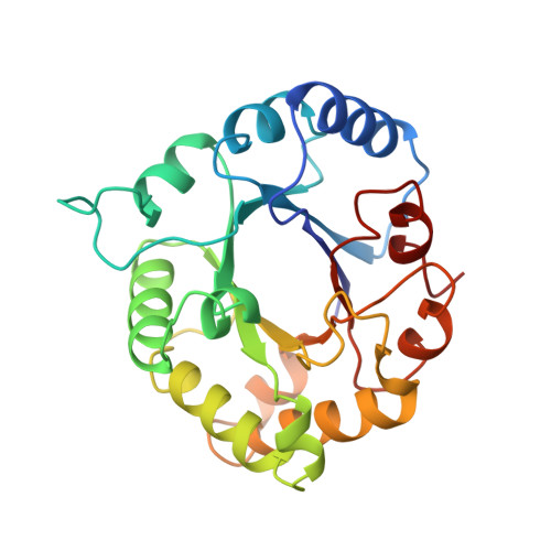
1TPW - PubMed Abstract:
The structural basis for the effect of the S96P mutation in chicken triosephosphate isomerase (cTIM) has been analyzed using a combination of X-ray crystallography and Fourier transform infrared spectroscopy. The X-ray structure is that of the enzyme complexed with phosphoglycolohydroxamate (PGH), an intermediate analogue, solved at a resolution of 1.9 A. The S96P mutation was identified as a second-site reverent when catalytically crippled mutants, E165D and H95N, were subjected to random mutagenesis. The presence of the second mutation leads to enhanced activity over the single mutation. However, the effect of the S96P mutation alone is to decrease the catalytic efficiency of the enzyme. The crystal structures of the S96P double mutants show that this bulky proline side chain alters the water structure within the active-site cavity (E165D; ref 1) and prevents nonproductive binding conformations of the substrate (H95N; ref 2). Comparison of the S96P single mutant structure with those of the wild-type cTIM, those of the single mutants (E165D and H95N), and those of the double mutants (E165D/S96P and H95N/S96P) begins to address the role of the conserved serine residue at this position. The results indicate that the residue positions the catalytic base E165 optimally for polarization of the substrate carbonyl, thereby aiding in proton abstraction. In addition, this residue is involved in positioning critical water molecules, thereby affecting the way in which water structure influences activity.
- Department of Biochemistry, Rosenstiel Basic Medical Sciences Research Center, Brandeis University, Waltham, Massachusetts 02254-9110, USA.
Organizational Affiliation: