The structure of the N-terminus of striated muscle alpha-tropomyosin in a chimeric peptide: nuclear magnetic resonance structure and circular dichroism studies.
Greenfield, N.J., Montelione, G.T., Farid, R.S., Hitchcock-DeGregori, S.E.(1998) Biochemistry 37: 7834-7843
- PubMed: 9601044 Search on PubMed
- DOI: https://doi.org/10.1021/bi973167m
- Primary Citation Related Structures:
1TMZ - PubMed Abstract:
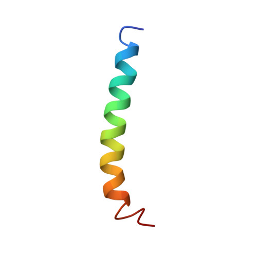
Tropomyosins (TMs) are highly conserved, coiled-coil, actin binding regulatory proteins found in most eukaryotic cells. The amino-terminal domain of 284-residue TMs is among the most conserved and functionally important regions. The first nine residues are proposed to bind to the carboxyl-terminal nine residues to form the "overlap" region between successive TMs, which bind along the actin filament. Here, the structure of the N-terminus of muscle alpha-TM, in a chimeric peptide, TMZip, has been solved using circular dichroism (CD) and two-dimensional proton nuclear magnetic resonance (2D 1H NMR) spectroscopy. Residues 1-14 of TMZip are the first 14 N-terminal residues of rabbit striated alpha-TM, and residues 15-32 of TMZip are the last 18 C-terminal residues of the yeast GCN4 transcription factor. CD measurements show that TMZip forms a two-stranded coiled-coil alpha-helix with an enthalpy of folding of -34 +/- 2 kcal/mol. In 2D1H NMR studies at 15 degrees C, pH 6.4, the peptide exhibits 123 sequential and medium range intrachain NOE cross peaks per chain, characteristic of alpha-helices extending from residue 1 to residue 29, together with 85 long-range NOE cross peaks arising from interchain interactions. The three-dimensional structure of TMZip has been determined using these data plus an additional 509 intrachain constraints per chain. The coiled-coil domain extends to the N-terminus. Amide hydrogen exchange studies, however, suggest that the TM region is less stable than the GCN4 region. The work reported here is the first atomic-resolution structure of any region of TM and it allows insight into the mechanism of the function of the highly conserved N-terminal domain.
- Department of Neuroscience and Cell Biology, Robert Wood Johnson Medical School, University of Medicine and Dentistry of New Jersey, Piscataway 08854-5635, USA.
Organizational Affiliation: