1TJF | pdb_00001tjf

The crystal structure of the N-terminal domain of CAP indicates variable oligomerisation


Experimental Data Snapshot

  • Method: X-RAY DIFFRACTION
  • Resolution: 2.21 Å
  • R-Value Free: 
    0.244 (Depositor), 0.241 (DCC) 
  • R-Value Work: 
    0.197 (Depositor), 0.195 (DCC) 
  • R-Value Observed: 
    0.197 (Depositor) 

Starting Model: experimental
View more details

wwPDB Validation 3D Report Full Report

Validation slider image for 1TJF

This is version 1.3 of the entry. See complete history

Literature

Structural evidence for variable oligomerization of the N-terminal domain of cyclase-associated protein (CAP).

Mohd Yusof, A.Hu, N.J.Wlodawer, A.Hofmann, A.

(2005) Proteins 58: 255-262

  • DOI: https://doi.org/10.1002/prot.20314
  • Primary Citation Related Structures: 
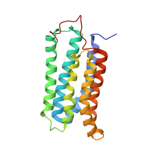
    1TJF

  • PubMed Abstract: 

    Cyclase-associated protein (CAP) is a highly conserved and widely distributed protein that links the nutritional response signaling to cytoskeleton remodeling. In yeast, CAP is a component of the adenylyl cyclase complex and helps to activate the Ras-mediated catalytic cycle of the cyclase. While the N-terminal domain of CAP (N-CAP) provides a binding site for adenylyl cyclase, the C-terminal domain (C-CAP) possesses actin binding activity. Our attempts to crystallize full-length recombinant CAP from Dictyostelium discoideum resulted in growth of orthorhombic crystals containing only the N-terminal domain (residues 42-227) due to auto-proteolytic cleavage. The structure was solved by molecular replacement with data at 2.2 A resolution. The present crystal structure allows the characterization of a head-to-tail N-CAP dimer in the asymmetric unit and a crystallographic side-to-side dimer. Comparison with previously published structures of N-CAP reveals variable modes of dimerization of this domain, but the presence of a common interface for the side-to-side dimer.


  • Organizational Affiliation
    • Institute of Structural & Molecular Biology, School of Biological Sciences, The University of Edinburgh, Scotland, United Kingdom.

Macromolecule Content 

  • Total Structure Weight: 41.5 kDa 
  • Atom Count: 3,169 
  • Modeled Residue Count: 371 
  • Deposited Residue Count: 372 
  • Unique protein chains: 1

Macromolecules

Find similar proteins by:|  3D Structure
Entity ID: 1
MoleculeChains  Sequence LengthOrganismDetailsImage
Adenylyl cyclase-associated protein
A, B
186Dictyostelium discoideumMutation(s): 0 
Gene Names: CAP
UniProt
Find proteins for P54654 (Dictyostelium discoideum)
Explore P54654 
Go to UniProtKB:  P54654
Entity Groups
Sequence Clusters30% Identity50% Identity70% Identity90% Identity95% Identity100% Identity
UniProt GroupP54654
Sequence Annotations
Expand
Reference Sequence

Experimental Data & Validation

Experimental Data

  • Method: X-RAY DIFFRACTION
  • Resolution: 2.21 Å
  • R-Value Free:  0.244 (Depositor), 0.241 (DCC) 
  • R-Value Work:  0.197 (Depositor), 0.195 (DCC) 
  • R-Value Observed: 0.197 (Depositor) 
Space Group: C 2 2 21
Unit Cell:
Length ( Å )Angle ( ˚ )
a = 71.24α = 90
b = 75.06β = 90
c = 162.874γ = 90
Software Package:
Software NamePurpose
CNSrefinement
MOSFLMdata reduction
CCP4data scaling

Structure Validation

View Full Validation Report


Entry History 

Deposition Data

Revision History  (Full details and data files)

  • Version 1.0: 2005-02-01
    Type: Initial release
  • Version 1.1: 2008-04-30
    Changes: Version format compliance
  • Version 1.2: 2011-07-13
    Changes: Version format compliance
  • Version 1.3: 2023-08-23
    Changes: Data collection, Database references, Derived calculations, Refinement description