1THX | pdb_00001thx

THIOREDOXIN-2


Experimental Data Snapshot

  • Method: X-RAY DIFFRACTION
  • Resolution: 1.60 Å
  • R-Value Free: 
    0.229 (Depositor), 0.180 (DCC) 
  • R-Value Work: 
    0.175 (Depositor), 0.172 (DCC) 
  • R-Value Observed: 
    0.175 (Depositor) 

wwPDB Validation 3D Report Full Report

Validation slider image for 1THX

This is version 1.4 of the entry. See complete history

Literature

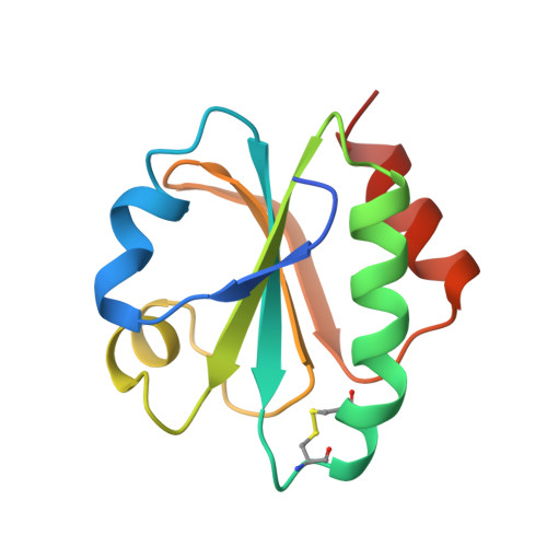
Crystal structure of thioredoxin-2 from Anabaena.

Saarinen, M.Gleason, F.K.Eklund, H.

(1995) Structure 3: 1097-1108

  • DOI: https://doi.org/10.1016/s0969-2126(01)00245-3
  • Primary Citation Related Structures: 
    1THX

  • PubMed Abstract: 

    Thioredoxins are ubiquitous proteins that serve as reducing agents and general protein disulfide reductases. The structures of thioredoxins from a number of species, including man and Escherichia coli, are known. Cyanobacteria, such as Anabaena, contain two thioredoxins that exhibit very different activities with target enzymes and share little sequence similarity. Thioredoxin-2 (Trx-2) from Anabaena resembles chloroplast type-f thioredoxin in its activities and the two proteins may be evolutionarily related. We have undertaken structural studies of Trx-2 in order to gain insights into the structure/function relationships of thioredoxins. Anabaena Trx-2, like E. coli thioredoxin, consists of a five-stranded beta sheet core surrounded by four alpha helices. The active site includes a conserved disulfide ring (in the sequence 31WCGPC35). An aspartate (E. coli) to tyrosine (Trx-2) substitution alters the position of this disulfide ring relative to the central pleated sheet. However, loss of this conserved aspartate does not affect the disulfide geometry. In the Trx-2 crystals, the N-terminal residues make extensive contacts with a symmetry-related molecule with hydrogen bonds to residues 74-76 mimicking thioredoxin-protein interactions. The overall three-dimensional structure of Trx-2 is similar to E. coli thioredoxin and other related disulfide oxido-reductases. Single amino acid substitutions around the protein interaction area probably account for the unusual enzymatic activities of Trx-2 and its ability to discriminate between substrate and non-substrate peptides.


  • Organizational Affiliation
    • Department of Molecular Biology, Swedish University of Agricultural Sciences, Uppsala, Sweden.

Macromolecule Content 

  • Total Structure Weight: 12.81 kDa 
  • Atom Count: 924 
  • Modeled Residue Count: 108 
  • Deposited Residue Count: 115 
  • Unique protein chains: 1

Macromolecules

Find similar proteins by:|  3D Structure
Entity ID: 1
MoleculeChains  Sequence LengthOrganismDetailsImage
THIOREDOXIN115Nostoc sp. PCC 7120 = FACHB-418Mutation(s): 0 
Gene Names: TRXA
UniProt
Find proteins for P20857 (Nostoc sp. (strain PCC 7120 / SAG 25.82 / UTEX 2576))
Explore P20857 
Go to UniProtKB:  P20857
Entity Groups
Sequence Clusters30% Identity50% Identity70% Identity90% Identity95% Identity100% Identity
UniProt GroupP20857
Sequence Annotations
Expand
Reference Sequence

Experimental Data & Validation

Experimental Data

  • Method: X-RAY DIFFRACTION
  • Resolution: 1.60 Å
  • R-Value Free:  0.229 (Depositor), 0.180 (DCC) 
  • R-Value Work:  0.175 (Depositor), 0.172 (DCC) 
  • R-Value Observed: 0.175 (Depositor) 
Space Group: P 21 21 21
Unit Cell:
Length ( Å )Angle ( ˚ )
a = 40.48α = 90
b = 39.78β = 90
c = 60.08γ = 90
Software Package:
Software NamePurpose
DENZO/CCP4data collection
X-PLORmodel building
X-PLORrefinement
DENZOdata reduction
CCP4data reduction
X-PLORphasing

Structure Validation

View Full Validation Report


Entry History 

Deposition Data

Revision History  (Full details and data files)

  • Version 1.0: 1995-10-15
    Type: Initial release
  • Version 1.1: 2008-03-03
    Changes: Version format compliance
  • Version 1.2: 2011-07-13
    Changes: Source and taxonomy, Version format compliance
  • Version 1.3: 2024-06-05
    Changes: Data collection, Database references, Other
  • Version 1.4: 2024-10-23
    Changes: Structure summary