1TGJ | pdb_00001tgj

HUMAN TRANSFORMING GROWTH FACTOR-BETA 3, CRYSTALLIZED FROM DIOXANE


Experimental Data Snapshot

  • Method: X-RAY DIFFRACTION
  • Resolution: 2.00 Å
  • R-Value Work: 
    0.175 (Depositor), 0.174 (DCC) 
  • R-Value Observed: 
    0.175 (Depositor) 

Starting Model: experimental
View more details

wwPDB Validation 3D Report Full Report

Validation slider image for 1TGJ

This is version 2.1 of the entry. See complete history

Literature

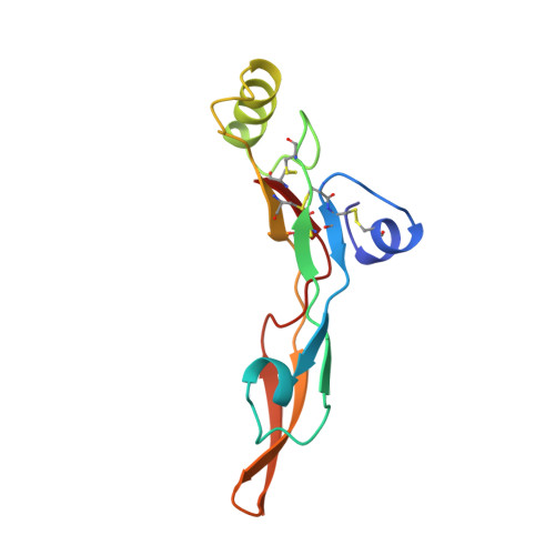
The crystal structure of TGF-beta 3 and comparison to TGF-beta 2: implications for receptor binding.

Mittl, P.R.Priestle, J.P.Cox, D.A.McMaster, G.Cerletti, N.Grutter, M.G.

(1996) Protein Sci 5: 1261-1271

  • DOI: https://doi.org/10.1002/pro.5560050705
  • Primary Citation Related Structures: 
    1TGJ, 1TGK

  • PubMed Abstract: 

    Transforming growth factors beta belong to a group of cytokines that control cellular proliferation and differentiation. Five isoforms are known that share approximately 75% sequence identity, but exert different biological activities. The structure of TGF-beta 3 was solved by X-ray crystallography and refined to a final R-factor of 17.5% at 2.0 A resolution. Comparison with the structure of TGF-beta 2 (Schlunegger MP, Grütter MG, 1992, Nature 358:430-434; Daopin S, Piez KA, Ogawa Y, Davies DR, 1992, Science 257:369-373) reveals a virtually identical central core. Differences exist in the conformations of the N-terminal alpha-helix and in the beta-sheet loops. In TGF-beta 3, the N-terminal alpha-helix has moved approximately 1 A away from the central core. This movement can be correlated with the mutation of Leu 17 to Val and Ala 47 to Pro in TGF-beta 3. The beta-sheet loops rotate as a rigid body 9 degrees around an axis that runs approximately parallel to the dimer axis. If these differences are recognized by the TGF-beta receptors, they might account for the individual cellular responses. A molecule of the precipitating agent dioxane is bound in a crystal contact, forming a hydrogen bond with Trp 32. This dioxane may occupy a carbohydrate-binding site, because dioxane possesses some structural similarity with a carbohydrate. The dioxane is in contact with two tryptophans, which are often involved in carbohydrate recognition.


  • Organizational Affiliation
    • Ciba-Geigy Ltd., Pharmaceutical Research, CH-4002 Basle, Switzerland.

Macromolecule Content 

  • Total Structure Weight: 12.82 kDa 
  • Atom Count: 981 
  • Modeled Residue Count: 112 
  • Deposited Residue Count: 112 
  • Unique protein chains: 1

Macromolecules

Find similar proteins by:|  3D Structure
Entity ID: 1
MoleculeChains  Sequence LengthOrganismDetailsImage
TRANSFORMING GROWTH FACTOR-BETA 3112Homo sapiensMutation(s): 0 
Gene Names: HTGF-BETA3
UniProt & NIH Common Fund Data Resources
Find proteins for P10600 (Homo sapiens)
Explore P10600 
Go to UniProtKB:  P10600
PHAROS:  P10600
GTEx:  ENSG00000119699 
Entity Groups
Sequence Clusters30% Identity50% Identity70% Identity90% Identity95% Identity100% Identity
UniProt GroupP10600
Sequence Annotations
Expand
Reference Sequence

Small Molecules

Ligands 1 Unique
IDChains Name / Formula / InChI Key2D Diagram3D Interactions
DIO

Query on DIO


Download:Ideal Coordinates CCD File
B [auth A]1,4-DIETHYLENE DIOXIDE
C4 H8 O2
RYHBNJHYFVUHQT-UHFFFAOYSA-N

Experimental Data & Validation

Experimental Data

  • Method: X-RAY DIFFRACTION
  • Resolution: 2.00 Å
  • R-Value Work:  0.175 (Depositor), 0.174 (DCC) 
  • R-Value Observed: 0.175 (Depositor) 
Space Group: P 32 2 1
Unit Cell:
Length ( Å )Angle ( ˚ )
a = 49.3α = 90
b = 49.3β = 90
c = 78.9γ = 120
Software Package:
Software NamePurpose
MADNESdata collection
ROTAVATAdata reduction
Agrovatadata reduction
X-PLORmodel building
X-PLORrefinement
MADNESdata reduction
CCP4data scaling
ROTAVATAdata scaling
X-PLORphasing

Structure Validation

View Full Validation Report


Entry History 

Deposition Data

Revision History  (Full details and data files)

  • Version 1.0: 1997-01-11
    Type: Initial release
  • Version 1.1: 2008-03-24
    Changes: Version format compliance
  • Version 1.2: 2011-07-13
    Changes: Derived calculations, Version format compliance
  • Version 1.3: 2018-04-18
    Changes: Data collection, Other, Refinement description
  • Version 2.0: 2024-04-03
    Changes: Atomic model, Data collection, Database references, Derived calculations, Refinement description
  • Version 2.1: 2024-10-23
    Changes: Structure summary