1TFG | pdb_00001tfg

AN UNUSUAL FEATURE REVEALED BY THE CRYSTAL STRUCTURE AT 2.2 ANGSTROMS RESOLUTION OF HUMAN TRANSFORMING GROWTH FACTOR-BETA2


Experimental Data Snapshot

  • Method: X-RAY DIFFRACTION
  • Resolution: 1.95 Å
  • R-Value Work: 
    0.194 (Depositor) 
  • R-Value Observed: 
    0.194 (Depositor) 

wwPDB Validation 3D Report Full Report

Validation slider image for 1TFG

This is version 1.5 of the entry. See complete history

Literature

An unusual feature revealed by the crystal structure at 2.2 A resolution of human transforming growth factor-beta 2.

Schlunegger, M.P.Grutter, M.G.

(1992) Nature 358: 430-434

  • DOI: https://doi.org/10.1038/358430a0
  • Primary Citation Related Structures: 
    1TFG

  • PubMed Abstract: 

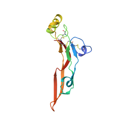
    Transforming growth factor type beta 2 (TGF-beta 2) is a member of an expanding family of growth factors that regulate proliferation and differentiation of many different cell types. TGF-beta 2 binds to various receptors, one of which was shown to be a serine/threonine kinase. TGF-beta 2 is involved in wound healing, bone formation and modulation of immune functions. We report here the crystal structure of TGF-beta 2 at 2.2 A resolution, which reveals a novel monomer fold and dimer association. The monomer consists of two antiparallel pairs of beta-strands forming a flat curved surface and a separate, long alpha-helix. The disulphide-rich core has one disulphide bone pointing through a ring formed by the sequence motifs Cys-Ala-Gly-Ala-Cys and Cys-Lys-Cys, which are themselves connected through the cysteines. Two monomers are connected through a single disulphide bridge and associate such that the helix of one subunit interacts with the concave beta-sheet surface of the other. Four exposed loop regions might determine receptor specificity. The structure provides a suitable model for the TGF-beta s and other members of the super-family and is the basis for the analysis of the TGF-beta 2 interactions with the receptor.


  • Organizational Affiliation
    • Department of Biotechnology, Pharmaceuticals Division, Ciba-Geigy, Basel, Switzerland.

Macromolecule Content 

  • Total Structure Weight: 12.73 kDa 
  • Atom Count: 974 
  • Modeled Residue Count: 112 
  • Deposited Residue Count: 112 
  • Unique protein chains: 1

Macromolecules

Find similar proteins by:|  3D Structure
Entity ID: 1
MoleculeChains  Sequence LengthOrganismDetailsImage
TRANSFORMING GROWTH FACTOR, BETA 2112Homo sapiensMutation(s): 0 
UniProt & NIH Common Fund Data Resources
Find proteins for P61812 (Homo sapiens)
Explore P61812 
Go to UniProtKB:  P61812
PHAROS:  P61812
GTEx:  ENSG00000092969 
Entity Groups
Sequence Clusters30% Identity50% Identity70% Identity90% Identity95% Identity100% Identity
UniProt GroupP61812
Sequence Annotations
Expand
Reference Sequence

Experimental Data & Validation

Experimental Data

  • Method: X-RAY DIFFRACTION
  • Resolution: 1.95 Å
  • R-Value Work:  0.194 (Depositor) 
  • R-Value Observed: 0.194 (Depositor) 
Space Group: P 32 2 1
Unit Cell:
Length ( Å )Angle ( ˚ )
a = 60.6α = 90
b = 60.6β = 90
c = 75.2γ = 120
Software Package:
Software NamePurpose
X-PLORmodel building
TNTrefinement
X-PLORrefinement
X-PLORphasing

Structure Validation

View Full Validation Report


Entry History 

Deposition Data

Revision History  (Full details and data files)

  • Version 1.0: 1993-10-31
    Type: Initial release
  • Version 1.1: 2008-03-24
    Changes: Version format compliance
  • Version 1.2: 2011-07-13
    Changes: Derived calculations, Version format compliance
  • Version 1.3: 2019-07-17
    Changes: Data collection, Other, Refinement description
  • Version 1.4: 2019-08-14
    Changes: Data collection, Refinement description
  • Version 1.5: 2024-11-20
    Changes: Data collection, Database references, Structure summary