1TBP

CRYSTAL STRUCTURE OF YEAST TATA-BINDING PROTEIN AND MODEL FOR INTERACTION WITH DNA


Experimental Data Snapshot

  • Method: X-RAY DIFFRACTION
  • Resolution: 2.60 Å
  • R-Value Work: 0.211 
  • R-Value Observed: 0.211 

wwPDB Validation   3D Report Full Report


This is version 1.3 of the entry. See complete history


Literature

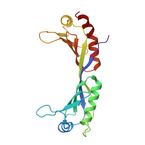
Crystal structure of yeast TATA-binding protein and model for interaction with DNA.

Chasman, D.I.Flaherty, K.M.Sharp, P.A.Kornberg, R.D.

(1993) Proc Natl Acad Sci U S A 90: 8174-8178

  • DOI: https://doi.org/10.1073/pnas.90.17.8174
  • Primary Citation of Related Structures:  
    1TBP

  • PubMed Abstract: 

    The C-terminal 179-aa region of yeast (Saccharomyces cerevisiae) TATA-binding protein (TBP), phylogenetically conserved and sufficient for many functions, formed crystals diffracting to 1.7-A resolution. The structure of the protein, determined by molecular replacement with coordinates from Arabidopsis TBP and refined to 2.6 A, differed from that in Arabidopsis slightly by an angle of about 12 degrees between two structurally nearly identical subdomains, indicative of a degree of conformational flexibility. A model for TBP-DNA interaction is proposed with the following important features: the long dimension of the protein follows the trajectory of the minor groove; two rows of basic residues conserved between the subdomains lie along the edges of the protein in proximity to the DNA phosphates; a band of hydrophobic residues runs down the middle of the groove; and amino acid residues whose mutation alters specificity for the second base of the TATA sequence are juxtaposed to that base.


  • Organizational Affiliation

    Center for Cancer Research, Massachusetts Institute of Technology, Cambridge 02139.


Macromolecules
Find similar proteins by:  (by identity cutoff)  |  3D Structure
Entity ID: 1
MoleculeChains Sequence LengthOrganismDetailsImage
TATA-BINDING PROTEIN
A, B
180Saccharomyces cerevisiaeMutation(s): 0 
UniProt
Find proteins for P13393 (Saccharomyces cerevisiae (strain ATCC 204508 / S288c))
Explore P13393 
Go to UniProtKB:  P13393
Entity Groups  
Sequence Clusters30% Identity50% Identity70% Identity90% Identity95% Identity100% Identity
UniProt GroupP13393
Sequence Annotations
Expand
  • Reference Sequence
Experimental Data & Validation

Experimental Data

  • Method: X-RAY DIFFRACTION
  • Resolution: 2.60 Å
  • R-Value Work: 0.211 
  • R-Value Observed: 0.211 
  • Space Group: P 31 2 1
Unit Cell:
Length ( Å )Angle ( ˚ )
a = 104.1α = 90
b = 104.1β = 90
c = 75.5γ = 120
Software Package:
Software NamePurpose
X-PLORmodel building
X-PLORrefinement
X-PLORphasing

Structure Validation

View Full Validation Report


Entry History 

Deposition Data

Revision History  (Full details and data files)

  • Version 1.0: 1994-01-31
    Type: Initial release
  • Version 1.1: 2008-03-24
    Changes: Version format compliance
  • Version 1.2: 2011-07-13
    Changes: Version format compliance
  • Version 1.3: 2024-02-14
    Changes: Data collection, Database references, Other