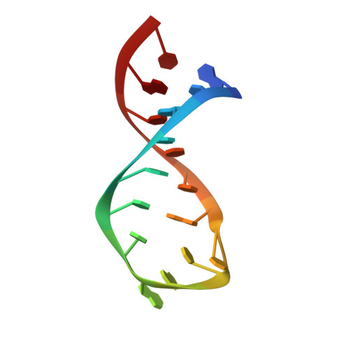
Nuclear Magnetic Resonance Structure of the Varkud Satellite Ribozyme Stem-Loop V RNA and Magnesium-Ion Binding from Chemical-Shift Mapping
Campbell, D.O., Legault, P.(2005) Biochemistry 44: 4157-4170
- PubMed: 15766243 Search on PubMed
- DOI: https://doi.org/10.1021/bi047963l
- Primary Citation Related Structures:
1TBK - PubMed Abstract:
An important step in the substrate recognition of the Neurospora Varkud Satellite (VS) ribozyme is the formation of a magnesium-dependent loop/loop interaction between the terminal loops of stem-loops I and V. We have studied the structure of stem-loop V by nuclear magnetic resonance spectroscopy and shown that it adopts a U-turn conformation, a common motif found in RNA. Structural comparisons indicate that the U-turn of stem-loop V fulfills some but not all of the structural characteristics found in canonical U-turn structures. This U-turn conformation exposes the Watson-Crick faces of the bases within stem-loop V (G697, A698, and C699) and makes them accessible for interaction with stem-loop I. Using chemical-shift mapping, we show that magnesium ions interact with the loop of the isolated stem-loop V and induce a conformational change that may be important for interaction with stem-loop I. This study expands our understanding of the role of U-turn motifs in RNA structure and function and provides insights into the mechanism of substrate recognition in the VS ribozyme.
- Department of Biochemistry and Molecular Biology, University of Georgia, Athens, Georgia 30602, USA.
Organizational Affiliation: