1T41 | pdb_00001t41

Crystal structure of human aldose reductase complexed with NADP and IDD552


Experimental Data Snapshot

  • Method: X-RAY DIFFRACTION
  • Resolution: 1.05 Å
  • R-Value Free: 
    0.133 (Depositor), 0.117 (DCC) 
  • R-Value Work: 
    0.108 (Depositor), 0.118 (DCC) 
  • R-Value Observed: 
    0.108 (Depositor) 

Starting Model: experimental
View more details

wwPDB Validation 3D Report Full Report

Validation slider image for 1T41

Ligand Structure Quality Assessment 


This is version 1.5 of the entry. See complete history

Literature

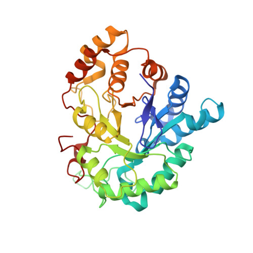
The crystallographic structure of the aldose reductase-IDD552 complex shows direct proton donation from tyrosine 48.

Ruiz, F.Hazemann, I.Mitschler, A.Joachimiak, A.Schneider, T.Karplus, M.Podjarny, A.

(2004) Acta Crystallogr D Biol Crystallogr 60: 1347-1354

  • DOI: https://doi.org/10.1107/S0907444904011370
  • Primary Citation Related Structures: 
    1T40, 1T41

  • PubMed Abstract: 

    The X-ray crystal structure of human aldose reductase (ALR2) in complex with the inhibitor IDD552 was determined using crystals obtained from two crystallization conditions with different pH values (pH 5 and 8). In both structures the charged carboxylic head of the inhibitor binds to the active site, making hydrogen-bond interactions with His110 and Tyr48 and electrostatic interactions with NADP+. There is an important difference between the two structures: the observation of a double conformation of the carboxylic acid moiety of the inhibitor at pH 8, with one water molecule interacting with the main configuration. This is the first time that a water molecule has been observed deep inside the ALR2 active site. Furthermore, in the configuration with the lower occupancy factor the difference electron-density map shows a clear peak (2.5sigma) for the H atom in the hydrogen bond between the inhibitor's carboxylic acid and the Tyr48 side-chain O atom. The position of this peak implies that this H atom is shared between both O atoms, indicating possible direct proton transfer from this residue to the inhibitor. This fact agrees with the model of the catalytic mechanism, in which the proton is donated by the Tyr48 hydroxyl to the substrate. These observations are useful both in drug design and in understanding the ALR2 mechanism.


  • Organizational Affiliation
    • UPR de Biologie Structurale, IGBMC, CNRS INSERM ULP, 1 Rue Laurent Fries, BP 163, 67404 Illkirch, France.

Macromolecule Content 

  • Total Structure Weight: 37.06 kDa 
  • Atom Count: 3,222 
  • Modeled Residue Count: 316 
  • Deposited Residue Count: 316 
  • Unique protein chains: 1

Macromolecules

Find similar proteins by:|  3D Structure
Entity ID: 1
MoleculeChains  Sequence LengthOrganismDetailsImage
Aldose reductase316Homo sapiensMutation(s): 0 
EC: 1.1.1.21 (PDB Primary Data), 1.1.1.372 (UniProt), 1.1.1.300 (UniProt), 1.1.1.54 (UniProt)
UniProt & NIH Common Fund Data Resources
Find proteins for P15121 (Homo sapiens)
Explore P15121 
Go to UniProtKB:  P15121
PHAROS:  P15121
GTEx:  ENSG00000085662 
Entity Groups
Sequence Clusters30% Identity50% Identity70% Identity90% Identity95% Identity100% Identity
UniProt GroupP15121
Sequence Annotations
Expand
Reference Sequence

Small Molecules

Ligands 2 Unique
IDChains Name / Formula / InChI Key2D Diagram3D Interactions
NAP

Query on NAP


Download:Ideal Coordinates CCD File
B [auth A]NADP NICOTINAMIDE-ADENINE-DINUCLEOTIDE PHOSPHATE
C21 H28 N7 O17 P3
XJLXINKUBYWONI-NNYOXOHSSA-N
ID5

Query on ID5


Download:Ideal Coordinates CCD File
C [auth A][5-FLUORO-2-({[(4,5,7-TRIFLUORO-1,3-BENZOTHIAZOL-2-YL)METHYL]AMINO}CARBONYL)PHENOXY]ACETIC ACID
C17 H10 F4 N2 O4 S
ZCAGEXZTORJQDZ-UHFFFAOYSA-N
Binding Affinity Annotations 
IDSourceBinding Affinity
ID5 BindingDB:  1T41 IC50: 11 (nM) from 1 assay(s)

Experimental Data & Validation

Experimental Data

  • Method: X-RAY DIFFRACTION
  • Resolution: 1.05 Å
  • R-Value Free:  0.133 (Depositor), 0.117 (DCC) 
  • R-Value Work:  0.108 (Depositor), 0.118 (DCC) 
  • R-Value Observed: 0.108 (Depositor) 
Space Group: P 1
Unit Cell:
Length ( Å )Angle ( ˚ )
a = 47.057α = 67.32
b = 47.525β = 76.79
c = 40.028γ = 75.82
Software Package:
Software NamePurpose
DENZOdata reduction
SCALEPACKdata scaling
X-PLORmodel building
SHELXL-97refinement
HKL-2000data reduction
X-PLORphasing

Structure Validation

View Full Validation Report


Ligand Structure Quality Assessment 


Entry History 

Deposition Data

Revision History  (Full details and data files)

  • Version 1.0: 2004-08-03
    Type: Initial release
  • Version 1.1: 2008-04-30
    Changes: Version format compliance
  • Version 1.2: 2011-07-13
    Changes: Version format compliance
  • Version 1.3: 2017-10-11
    Changes: Refinement description
  • Version 1.4: 2024-03-13
    Changes: Data collection, Database references, Derived calculations
  • Version 1.5: 2024-04-03
    Changes: Refinement description