1T3B | pdb_00001t3b

X-ray Structure of DsbC from Haemophilus influenzae


Experimental Data Snapshot

  • Method: X-RAY DIFFRACTION
  • Resolution: 2.50 Å
  • R-Value Free: 
    0.295 (Depositor), 0.305 (DCC) 
  • R-Value Work: 
    0.246 (Depositor), 0.255 (DCC) 
  • R-Value Observed: 
    0.249 (Depositor) 

Starting Model: experimental
View more details

wwPDB Validation 3D Report Full Report

Validation slider image for 1T3B

This is version 1.4 of the entry. See complete history

Literature

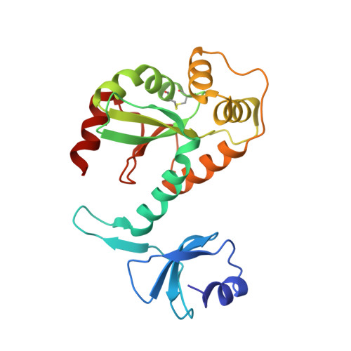
Structure of DsbC from Haemophilus influenzae.

Zhang, M.Monzingo, A.F.Segatori, L.Georgiou, G.Robertus, J.D.

(2004) Acta Crystallogr D Biol Crystallogr 60: 1512-1518

  • DOI: https://doi.org/10.1107/S0907444904014593
  • Primary Citation Related Structures: 
    1T3B

  • PubMed Abstract: 

    Bacterial DsbC proteins are involved in rearranging or reducing mismatched disulfide bonds folding within the periplasm. The X-ray structure of the enzyme from Haemophilus influenzae has been solved and compared with the known structure of the Escherichia coli protein. The proteins act as V-shaped dimers with a large cleft to accommodate substrate proteins. The dimers are anchored by a small N-terminal domain, but have a flexible linker region which allows the larger C-terminal domain, with its reactive sulfhydryls, to clamp down on substrates. The overall folds are very similar, but the comparison shows a wider range of hinge motions than previously thought. The crystal packing of the H. influenzae protein allows the movement of the N-terminal domain with respect to the C-terminal domain through motions in the flexible hinge, generating high thermal parameters and unusually high anisotropy in the crystallographic data.


  • Organizational Affiliation
    • Institute for Cellular and Molecular Biology, Department of Chemistry and Biochemistry, 1 University Station, University of Texas at Austin, Austin, TX 78712, USA.

Macromolecule Content 

  • Total Structure Weight: 23.53 kDa 
  • Atom Count: 1,574 
  • Modeled Residue Count: 209 
  • Deposited Residue Count: 211 
  • Unique protein chains: 1

Macromolecules

Find similar proteins by:|  3D Structure
Entity ID: 1
MoleculeChains  Sequence LengthOrganismDetailsImage
Thiol:disulfide interchange protein dsbC211Haemophilus influenzaeMutation(s): 0 
Gene Names: DSBCXPRAHI1213
EC: 5.3.4.1
UniProt
Find proteins for P45111 (Haemophilus influenzae (strain ATCC 51907 / DSM 11121 / KW20 / Rd))
Explore P45111 
Go to UniProtKB:  P45111
Entity Groups
Sequence Clusters30% Identity50% Identity70% Identity90% Identity95% Identity100% Identity
UniProt GroupP45111
Sequence Annotations
Expand
Reference Sequence

Experimental Data & Validation

Experimental Data

  • Method: X-RAY DIFFRACTION
  • Resolution: 2.50 Å
  • R-Value Free:  0.295 (Depositor), 0.305 (DCC) 
  • R-Value Work:  0.246 (Depositor), 0.255 (DCC) 
  • R-Value Observed: 0.249 (Depositor) 
Space Group: P 43 21 2
Unit Cell:
Length ( Å )Angle ( ˚ )
a = 74.92α = 90
b = 74.92β = 90
c = 103.38γ = 90
Software Package:
Software NamePurpose
DENZOdata reduction
SCALEPACKdata scaling
EPMRphasing
CNSrefinement

Structure Validation

View Full Validation Report


Entry History 

Deposition Data

Revision History  (Full details and data files)

  • Version 1.0: 2004-09-07
    Type: Initial release
  • Version 1.1: 2008-04-30
    Changes: Version format compliance
  • Version 1.2: 2011-07-13
    Changes: Version format compliance
  • Version 1.3: 2023-08-23
    Changes: Data collection, Database references, Refinement description
  • Version 1.4: 2024-10-30
    Changes: Structure summary