1T0J

Crystal structure of a complex between voltage-gated calcium channel beta2a subunit and a peptide of the alpha1c subunit


Experimental Data Snapshot

  • Method: X-RAY DIFFRACTION
  • Resolution: 2.00 Å
  • R-Value Free: 0.242 
  • R-Value Work: 0.200 
  • R-Value Observed: 0.202 

wwPDB Validation   3D Report Full Report


This is version 1.2 of the entry. See complete history


Literature

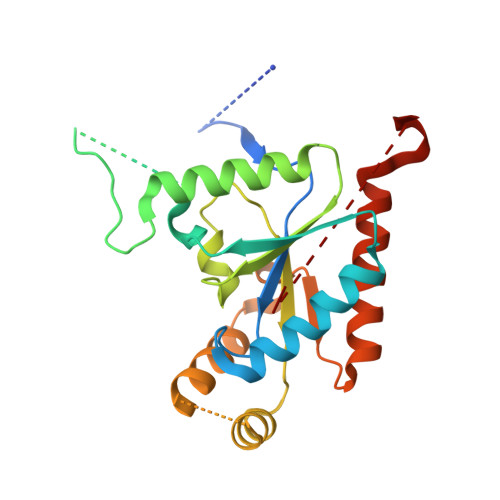
Structure of a complex between a voltage-gated calcium channel beta-subunit and an alpha-subunit domain.

Van Petegem, F.Clark, K.A.Chatelain, F.C.Minor, D.L.

(2004) Nature 429: 671-675

  • DOI: 10.1038/nature02588
  • Primary Citation of Related Structures:  
    1T0H, 1T0J

  • PubMed Abstract: 
  • Voltage-gated calcium channels (Ca(V)s) govern muscle contraction, hormone and neurotransmitter release, neuronal migration, activation of calcium-dependent signalling cascades, and synaptic input integration. An essential Ca(V) intracellular protein, the beta-subunit (Ca(V)beta), binds a conserved domain (the alpha-interaction domain, AID) between transmembrane domains I and II of the pore-forming alpha(1) subunit and profoundly affects multiple channel properties such as voltage-dependent activation, inactivation rates, G-protein modulation, drug sensitivity and cell surface expression ...

    Voltage-gated calcium channels (Ca(V)s) govern muscle contraction, hormone and neurotransmitter release, neuronal migration, activation of calcium-dependent signalling cascades, and synaptic input integration. An essential Ca(V) intracellular protein, the beta-subunit (Ca(V)beta), binds a conserved domain (the alpha-interaction domain, AID) between transmembrane domains I and II of the pore-forming alpha(1) subunit and profoundly affects multiple channel properties such as voltage-dependent activation, inactivation rates, G-protein modulation, drug sensitivity and cell surface expression. Here, we report the high-resolution crystal structures of the Ca(V)beta2a conserved core, alone and in complex with the AID. Previous work suggested that a conserved region, the beta-interaction domain (BID), formed the AID-binding site; however, this region is largely buried in the Ca(V)beta core and is unavailable for protein-protein interactions. The structure of the AID-Ca(V)beta2a complex shows instead that Ca(V)beta2a engages the AID through an extensive, conserved hydrophobic cleft (named the alpha-binding pocket, ABP). The ABP-AID interaction positions one end of the Ca(V)beta near the intracellular end of a pore-lining segment, called IS6, that has a critical role in Ca(V) inactivation. Together, these data suggest that Ca(V)betas influence Ca(V) gating by direct modulation of IS6 movement within the channel pore.


    Organizational Affiliation

    Cardiovascular Research Institute, Department of Biochemistry and Biophysics, University of California San Francisco, 513 Parnassus Avenue, Box 0130, San Francisco, California 94143, USA.


Macromolecules
Find similar proteins by:  (by identity cutoff)  |  3D Structure
Entity ID: 1
MoleculeChainsSequence LengthOrganismDetailsImage
voltage-gated calcium channel subunit beta2aA132Rattus norvegicusMutation(s): 0 
Gene Names: CACNB2ACacnb2Cacnlb2
UniProt
Find proteins for Q8VGC3 (Rattus norvegicus)
Explore Q8VGC3 
Go to UniProtKB:  Q8VGC3
Entity Groups  
Sequence Clusters30% Identity50% Identity70% Identity90% Identity95% Identity100% Identity
UniProt GroupQ8VGC3
Protein Feature View
Expand
  • Reference Sequence
Find similar proteins by:  (by identity cutoff)  |  3D Structure
Entity ID: 2
MoleculeChainsSequence LengthOrganismDetailsImage
voltage-gated calcium channel subunit beta2aB224Rattus norvegicusMutation(s): 0 
Gene Names: CACNB2ACacnb2Cacnlb2
UniProt
Find proteins for Q8VGC3 (Rattus norvegicus)
Explore Q8VGC3 
Go to UniProtKB:  Q8VGC3
Entity Groups  
Sequence Clusters30% Identity50% Identity70% Identity90% Identity95% Identity100% Identity
UniProt GroupQ8VGC3
Protein Feature View
Expand
  • Reference Sequence

Find similar proteins by:  Sequence   |   3D Structure  

Entity ID: 3
MoleculeChainsSequence LengthOrganismDetailsImage
Voltage-dependent L-type calcium channel alpha-1C subunitC20Homo sapiensMutation(s): 0 
Gene Names: CACNA1CCACNL1A1CCHL1A1CACH2CACN2
UniProt & NIH Common Fund Data Resources
Find proteins for Q13936 (Homo sapiens)
Explore Q13936 
Go to UniProtKB:  Q13936
PHAROS:  Q13936
Entity Groups  
Sequence Clusters30% Identity50% Identity70% Identity90% Identity95% Identity100% Identity
UniProt GroupQ13936
Protein Feature View
Expand
  • Reference Sequence
Small Molecules
Ligands 1 Unique
IDChainsName / Formula / InChI Key2D Diagram3D Interactions
CL
Query on CL

Download Ideal Coordinates CCD File 
D [auth B]CHLORIDE ION
Cl
VEXZGXHMUGYJMC-UHFFFAOYSA-M
 Ligand Interaction
Experimental Data & Validation

Experimental Data

  • Method: X-RAY DIFFRACTION
  • Resolution: 2.00 Å
  • R-Value Free: 0.242 
  • R-Value Work: 0.200 
  • R-Value Observed: 0.202 
  • Space Group: P 1
Unit Cell:
Length ( Å )Angle ( ˚ )
a = 36.756α = 96.81
b = 45.33β = 102.46
c = 60.652γ = 98.53
Software Package:
Software NamePurpose
DENZOdata reduction
SCALEPACKdata scaling
AMoREphasing
REFMACrefinement

Structure Validation

View Full Validation Report


Entry History 

Deposition Data

Revision History  (Full details and data files)

  • Version 1.0: 2004-06-15
    Type: Initial release
  • Version 1.1: 2008-04-30
    Changes: Version format compliance
  • Version 1.2: 2011-07-13
    Changes: Advisory, Version format compliance