1T0C | pdb_00001t0c

Solution Structure of Human Proinsulin C-Peptide


Experimental Data Snapshot

  • Method: SOLUTION NMR
  • Conformers Calculated: 800 
  • Conformers Submitted: 30 
  • Selection Criteria: structures with the lowest energy 

wwPDB Validation 3D Report Full Report

Validation slider image for 1T0C

This is version 1.4 of the entry. See complete history

Literature

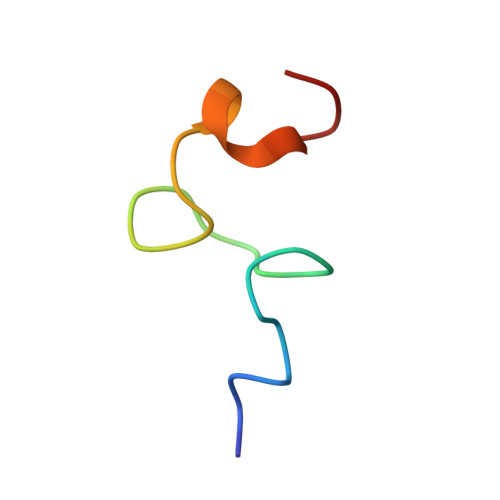
Solution structure of human proinsulin C-peptide

Munte, C.E.Vilela, L.Kalbitzer, H.-R.Garratt, R.C.

(2005) FEBS J 272: 4284-4293

  • DOI: https://doi.org/10.1111/j.1742-4658.2005.04843.x
  • Primary Citation Related Structures: 
    1T0C

  • PubMed Abstract: 

    The C-peptide of proinsulin is important for the biosynthesis of insulin, but has been considered for a long time to be biologically inert. Recent studies in diabetic patients have stimulated a new debate about its possible regulatory role, suggesting that it is a hormonally active peptide. We describe structural studies of the C-peptide using 2D NMR spectroscopy. In aqueous solution, the NOE patterns and chemical shifts indicate that the ensemble is a nonrandom structure and contains substructures with defined local conformations. These are more clearly visible in 50% H2O/50% 2,2,2-trifluoroethanol. The N-terminal region (residues 2-5) forms a type I beta-turn, whereas the C-terminal region (residues 27-31) presents the most well-defined structure of the whole molecule including a type III'beta-turn. The C-terminal pentapeptide (EGSLQ) has been suggested to be responsible for chiral interactions with an as yet uncharacterized, probably a G-protein-coupled, receptor. The three central regions of the molecule (residues 9-12, 15-18 and 22-25) show tendencies to form beta-bends. We propose that the structure described here for the C-terminal pentapeptide is consistent with the previously postulated CA knuckle, believed to represent the active site of the C-peptide of human proinsulin.


  • Organizational Affiliation
    • Instituto de Física de São Carlos, Universidade de São Paulo, São Carlos, Brazil.

Macromolecule Content 

  • Total Structure Weight: 3.02 kDa 
  • Atom Count: 212 
  • Modeled Residue Count: 31 
  • Deposited Residue Count: 31 
  • Unique protein chains: 1

Macromolecules

Find similar proteins by:|  3D Structure
Entity ID: 1
MoleculeChains  Sequence LengthOrganismDetailsImage
Insulin31Homo sapiensMutation(s): 0 
Gene Names: INS
UniProt & NIH Common Fund Data Resources
Find proteins for P01308 (Homo sapiens)
Explore P01308 
Go to UniProtKB:  P01308
PHAROS:  P01308
GTEx:  ENSG00000254647 
Entity Groups
Sequence Clusters30% Identity50% Identity70% Identity90% Identity95% Identity100% Identity
UniProt GroupP01308
Sequence Annotations
Expand
Reference Sequence

Experimental Data & Validation

Experimental Data

  • Method: SOLUTION NMR
  • Conformers Calculated: 800 
  • Conformers Submitted: 30 
  • Selection Criteria: structures with the lowest energy 

Structure Validation

View Full Validation Report


Entry History 

Deposition Data

Revision History  (Full details and data files)

  • Version 1.0: 2005-08-16
    Type: Initial release
  • Version 1.1: 2008-04-30
    Changes: Version format compliance
  • Version 1.2: 2011-07-13
    Changes: Version format compliance
  • Version 1.3: 2022-03-02
    Changes: Data collection, Database references, Derived calculations
  • Version 1.4: 2024-05-22
    Changes: Data collection