Transfer of a beta-turn structure to a new protein context.
Hynes, T.R., Kautz, R.A., Goodman, M.A., Gill, J.F., Fox, R.O.(1989) Nature 339: 73-76
- PubMed: 2716830 Search on PubMed
- DOI: https://doi.org/10.1038/339073a0
- Primary Citation Related Structures:
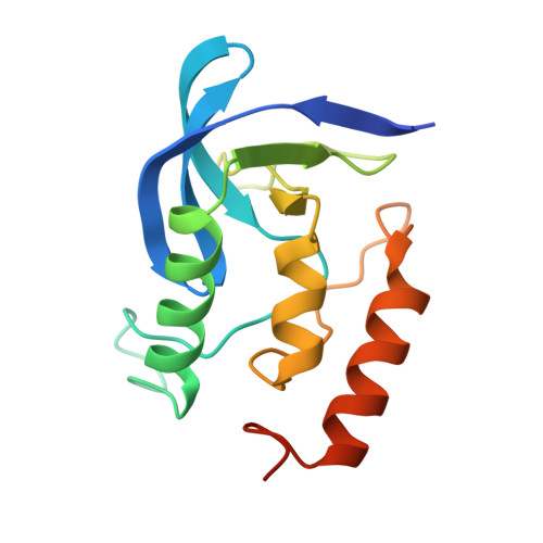
1SYB - PubMed Abstract:
Four-residue beta-turns and larger loop structures represent a significant fraction of globular protein surfaces and play an important role in determining the conformation and specificity of enzyme active sites and antibody-combining sites. Turns are an attractive starting point to develop protein design methods, as they involve a small number of consecutive residues, adopt a limited number of defined conformations and are minimally constrained by packing interactions with the remainder of the protein. The ability to substitute one beta-turn geometry for another will extend protein engineering beyond the redecoration of fixed backbone conformations to include local restructuring and the repositioning of surface side chains. To determine the feasibility and to examine the effect of such a structural modification on the fold and thermodynamic stability of a globular protein, we have substituted a five-residue turn sequence from concanavalin A for a type I' beta-turn in staphylococcal nuclease. The resulting hybrid protein is folded and has full nuclease enzymatic activity but reduced thermodynamic stability. The crystal structure of the hybrid protein reveals that the guest turn sequence retains the conformation of the parent concanavalin A structure when substituted in the nuclease host.
- Department of Molecular Biophysics and Biochemistry, Yale University, New Haven, Connecticut 06511.
Organizational Affiliation: