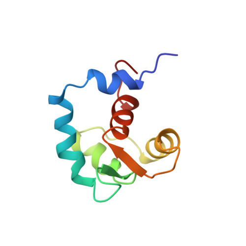
Calcium-induced structural transition in the regulatory domain of human cardiac troponin C.
Spyracopoulos, L., Li, M.X., Sia, S.K., Gagne, S.M., Chandra, M., Solaro, R.J., Sykes, B.D.(1997) Biochemistry 36: 12138-12146
- PubMed: 9315850 Search on PubMed
- DOI: https://doi.org/10.1021/bi971223d
- Primary Citation Related Structures:
1AP4, 1SPY - PubMed Abstract:
While calcium binding to troponin C (TnC) triggers the contraction of both skeletal and cardiac muscle, there is clear evidence that different mechanisms may be involved. For example, activation of heart myofilaments occurs with binding to a single regulatory site on TnC, whereas activation of fast skeletal myofilaments occurs with binding to two regulatory sites. The physiological difference between activation of cardiac and skeletal myofilaments is not understood at the molecular level due to a lack of structural details for the response of cardiac TnC to calcium. We determined the solution structures of the apo and calcium-saturated regulatory domain of human cardiac TnC by using multinuclear, multidimensional nuclear magnetic resonance spectroscopy. The structure of apo human cardiac TnC is very similar to that of apo turkey skeletal TnC even though there are critical amino acid substitutions in site I. In contrast to the case with the skeletal protein, the calcium-induced conformational transition in the cardiac regulatory domain does not involve an "opening" of the regulatory domain, and the concomitant exposure of a substantial hydrophobic surface area. This result has important implications with regard to potential unique aspects of the interaction of cardiac TnC with cardiac troponin I and of modification of cardiac myofilament regulation by calcium-sensitizer drugs.
- MRC Group in Protein Structure and Function, Department of Biochemistry, University of Alberta, Edmonton, Canada.
Organizational Affiliation: