1SN6 | pdb_00001sn6

NMR solution structure of human Saposin C in SDS micelles


Experimental Data Snapshot

  • Method: SOLUTION NMR
  • Conformers Calculated: 200 
  • Conformers Submitted: 20 
  • Selection Criteria: structures with the lowest energy 

wwPDB Validation 3D Report Full Report

Validation slider image for 1SN6

This is version 1.4 of the entry. See complete history

Literature

Solution structure of human saposin C in a detergent environment.

Hawkins, C.A.Alba, E.Tjandra, N.

(2005) J Mol Biology 346: 1381-1392

  • DOI: https://doi.org/10.1016/j.jmb.2004.12.045
  • Primary Citation Related Structures: 
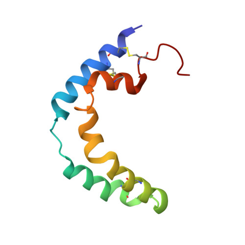
    1SN6

  • PubMed Abstract: 

    Saposin C is a lysosomal, membrane-binding protein that acts as an activator for the hydrolysis of glucosylceramide by the enzyme glucocerebrosidase. We used high-resolution NMR to determine the three-dimensional solution structure of saposin C in the presence of the detergent sodium dodecyl sulfate (SDS). This structure provides the first representation of membrane bound saposin C at the atomic level. In the presence of SDS, the protein adopts an open conformation with an exposed hydrophobic pocket. In contrast, the previously reported NMR structure of saposin C in the absence of SDS is compact and contains a hydrophobic core that is not exposed to the solvent. NMR data indicate that the SDS molecules interact with the hydrophobic pocket. The structure of saposin C in the presence of SDS is very similar to a monomer in the saposin B homodimer structure. Their comparison reveals possible similarity in the type of protein/lipid interaction as well as structural components differentiating their quaternary structures and functional specificity.


  • Organizational Affiliation
    • Laboratory of Biophysical Chemistry, National Heart, Lung, and Blood Institute, National Institutes of Health, 50 Center Drive, Bethesda, MD 20892, USA.

Macromolecule Content 

  • Total Structure Weight: 9.42 kDa 
  • Atom Count: 653 
  • Modeled Residue Count: 84 
  • Deposited Residue Count: 84 
  • Unique protein chains: 1

Macromolecules

Find similar proteins by:|  3D Structure
Entity ID: 1
MoleculeChains  Sequence LengthOrganismDetailsImage
Proactivator polypeptide84Homo sapiensMutation(s): 0 
Gene Names: PSAP
Membrane Entity: Yes 
UniProt & NIH Common Fund Data Resources
Find proteins for P07602 (Homo sapiens)
Explore P07602 
Go to UniProtKB:  P07602
PHAROS:  P07602
GTEx:  ENSG00000197746 
Entity Groups
Sequence Clusters30% Identity50% Identity70% Identity90% Identity95% Identity100% Identity
UniProt GroupP07602
Sequence Annotations
Expand
Reference Sequence

Experimental Data & Validation

Experimental Data

  • Method: SOLUTION NMR
  • Conformers Calculated: 200 
  • Conformers Submitted: 20 
  • Selection Criteria: structures with the lowest energy 

Structure Validation

View Full Validation Report


Entry History 

Deposition Data

Revision History  (Full details and data files)

  • Version 1.0: 2005-03-01
    Type: Initial release
  • Version 1.1: 2008-04-29
    Changes: Version format compliance
  • Version 1.2: 2011-07-13
    Changes: Version format compliance
  • Version 1.3: 2022-03-02
    Changes: Data collection, Database references, Derived calculations
  • Version 1.4: 2024-11-13
    Changes: Data collection, Structure summary