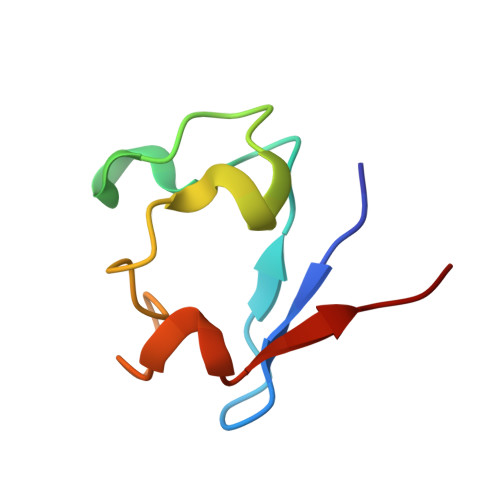
The unique hydrogen bonded water in the reduced form of Clostridium pasteurianum rubredoxin and its possible role in electron transfer
Park, I.Y., Youn, B., Harley, J.L., Eidsness, M.K., Smith, E., Ichiye, T., Kang, C.(2004) J Biol Inorg Chem 9: 423-428
- PubMed: 15067525 Search on PubMed
- DOI: https://doi.org/10.1007/s00775-004-0542-3
- Primary Citation Related Structures:
1SMM, 1SMU, 1SMW - PubMed Abstract:
Rubredoxin is a small iron-sulfur (FeS4) protein involved in oxidation-reduction reactions. The side chain of Leu41 near the iron-sulfur center has two conformations, which we suggested previously serve as a gate for a water molecule during the electron transfer process. To establish the role of residue 41 in electron transfer, an [L41A] mutant of Clostridium pasteurianum rubredoxin was constructed and crystallized in both oxidation states. Despite the lack of the gating side chain in this protein, the structure of the reduced [L41A] rubredoxin reveals a specific water molecule in the same position as observed in the reduced wild-type rubredoxin. In contrast, both the wild-type and [L41A] rubredoxins in the oxidized state do not have water molecules in this location. The reduction potential of the [L41A] variant was approximately 50 mV more positive than wild-type. Based on these observations, it is proposed that the site around the Sgamma of Cys9 serves as a port for an electron acceptor. Lastly, the Fe-S distances of the reduced rubredoxin are expanded, while the hydrogen bonds between Sgamma of the cysteines and the backbone amide nitrogens are shortened compared to its oxidized counterpart. This small structural perturbation in the Fe(II)/Fe(III) transition is closely related to the small energy difference which is important in an effective electron transfer agent.
- School of Molecular Biosciences, Washington State University, Pullman, WA 99164-4660, USA.
Organizational Affiliation: