1SMD | pdb_00001smd

HUMAN SALIVARY AMYLASE


Experimental Data Snapshot

  • Method: X-RAY DIFFRACTION
  • Resolution: 1.60 Å
  • R-Value Work: 
    0.184 (Depositor), 0.194 (DCC) 

wwPDB Validation 3D Report Full Report

Validation slider image for 1SMD

This is version 2.1 of the entry. See complete history

Literature

Structure of human salivary alpha-amylase at 1.6 A resolution: implications for its role in the oral cavity.

Ramasubbu, N.Paloth, V.Luo, Y.Brayer, G.D.Levine, M.J.

(1996) Acta Crystallogr D Biol Crystallogr 52: 435-446

  • DOI: https://doi.org/10.1107/S0907444995014119
  • Primary Citation Related Structures: 
    1SMD

  • PubMed Abstract: 

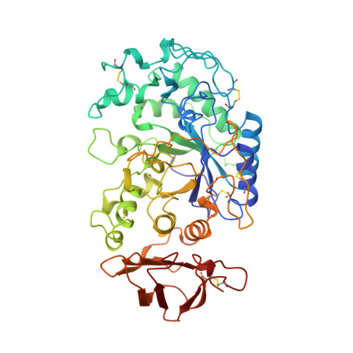
    Salivary alpha-amylase, a major component of human saliva, plays a role in the initial digestion of starch and may be involved in the colonization of bacteria involved in early dental plaque formation. The three-dimensional atomic structure of salivary amylase has been determined to understand the structure-function relationships of this enzyme. This structure was refined to an R value of 18.4% with 496 amino-acid residues, one calcium ion, one chloride ion and 170 water molecules. Salivary amylase folds into a multidomain structure consisting of three domains, A, B and C. Domain A has a (beta/alpha)(8-) barrel structure, domain B has no definite topology and domain C has a Greek-key barrel structure. The Ca(2+) ion is bound to Asnl00, Arg158, Asp167, His201 and three water molecules. The Cl(-) ion is bound to Arg195, Asn298 and Arg337 and one water molecule. The highly mobile glycine-rich loop 304-310 may act as a gateway for substrate binding and be involved in a 'trap-release' mechanism in the hydrolysis of substrates. Strategic placement of calcium and chloride ions, as well as histidine and tryptophan residues may play a role in differentiating between the glycone and aglycone ends of the polysaccharide substrates. Salivary amylase also possesses a suitable site for binding to enamel surfaces and provides potential sites for the binding of bacterial adhesins.


  • Organizational Affiliation
    • Department of Oral Biology and Dental Research Institute, School of Dental Medicine, State University of New York at Buffalo, BY 14214, USA. subbu@crystal1.sdm.buffalo.edu

Macromolecule Content 

  • Total Structure Weight: 56.03 kDa 
  • Atom Count: 4,117 
  • Modeled Residue Count: 496 
  • Deposited Residue Count: 496 
  • Unique protein chains: 1

Macromolecules

Find similar proteins by:|  3D Structure
Entity ID: 1
MoleculeChains  Sequence LengthOrganismDetailsImage
AMYLASE496Homo sapiensMutation(s): 0 
EC: 3.2.1.1
UniProt & NIH Common Fund Data Resources
Find proteins for P0DUB6 (Homo sapiens)
Explore P0DUB6 
Go to UniProtKB:  P0DUB6
GTEx:  ENSG00000237763 
Entity Groups
Sequence Clusters30% Identity50% Identity70% Identity90% Identity95% Identity100% Identity
UniProt GroupP0DUB6
Sequence Annotations
Expand
Reference Sequence

Small Molecules

Modified Residues  1 Unique
IDChains TypeFormula2D DiagramParent
PCA
Query on PCA
A
L-PEPTIDE LINKINGC5 H7 N O3GLN

Experimental Data & Validation

Experimental Data

  • Method: X-RAY DIFFRACTION
  • Resolution: 1.60 Å
  • R-Value Work:  0.184 (Depositor), 0.194 (DCC) 
Space Group: P 21 21 21
Unit Cell:
Length ( Å )Angle ( ˚ )
a = 53.2α = 90
b = 75.4β = 90
c = 136.6γ = 90
Software Package:
Software NamePurpose
X-PLORmodel building
X-PLORrefinement
X-PLORphasing

Structure Validation

View Full Validation Report


Entry History 

Deposition Data

Revision History  (Full details and data files)

  • Version 1.0: 1996-07-11
    Type: Initial release
  • Version 1.1: 2008-03-24
    Changes: Version format compliance
  • Version 1.2: 2011-07-13
    Changes: Version format compliance
  • Version 2.0: 2019-12-25
    Changes: Derived calculations, Other, Polymer sequence
  • Version 2.1: 2024-10-16
    Changes: Data collection, Database references, Derived calculations, Structure summary