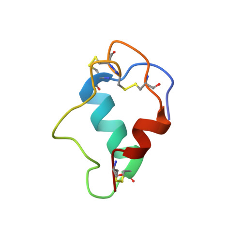
Mini-proinsulin and mini-IGF-I: homologous protein sequences encoding non-homologous structures.
Hua, Q.X., Hu, S.Q., Jia, W., Chu, Y.C., Burke, G.T., Wang, S.H., Wang, R.Y., Katsoyannis, P.G., Weiss, M.A.(1998) J Mol Biology 277: 103-118
- PubMed: 9514738 Search on PubMed
- DOI: https://doi.org/10.1006/jmbi.1997.1574
- Primary Citation Related Structures:
1SJT, 1SJU - PubMed Abstract:
Protein minimization highlights essential determinants of structure and function. Minimal models of proinsulin and insulin-like growth factor I contain homologous A and B domains as single-chain analogues. Such models (designated mini-proinsulin and mini-IGF-I) have attracted wide interest due to their native foldability but complete absence of biological activity. The crystal structure of mini-proinsulin, determined as a T3R3 hexamer, is similar to that of the native insulin hexamer. Here, we describe the solution structure of a monomeric mini-proinsulin under physiologic conditions and compare this structure to that of the corresponding two-chain analogue. The two proteins each contain substitutions in the B-chain (HisB10-->Asp and ProB28-->Asp) designed to destabilize self-association by electrostatic repulsion; the proteins differ by the presence or absence of a peptide bond between LysB29 and GlyA1. The structures are essentially identical, resembling in each case the T-state crystallographic protomer. Differences are observed near the site of cross-linking: the adjoining A1-A8 alpha-helix (variable among crystal structures) is less well-ordered in mini-proinsulin than in the two-chain variant. The single-chain analogue is not completely inactive: its affinity for the insulin receptor is 1500-fold lower than that of the two-chain analogue. Moreover, at saturating concentrations mini-proinsulin retains the ability to stimulate lipogenesis in adipocytes (native biological potency). These results suggest that a change in the conformation of insulin, as tethered by the B29-A1 peptide bond, optimizes affinity but is not integral to the mechanism of transmembrane signaling. Surprisingly, the tertiary structure of mini-proinsulin differs from that of mini-IGF-I (main-chain rms deviation 4.5 A) despite strict conservation of non-polar residues in their respective hydrophobic cores (side-chain rms deviation 4.9 A). Three-dimensional profile scores suggest that the two structures each provide acceptable templates for threading of insulin-like sequences. Mini-proinsulin and mini-IGF-I thus provide examples of homologous protein sequences encoding non-homologous structures.
- Center for Molecular Oncology and Department of Biochemistry and Molecular Biology, The University of Chicago, Chicago, IL 60637, USA.
Organizational Affiliation: