1SEI | pdb_00001sei

STRUCTURE OF 30S RIBOSOMAL PROTEIN S8


Experimental Data Snapshot

  • Method: X-RAY DIFFRACTION
  • Resolution: 1.90 Å
  • R-Value Free: 
    0.288 (Depositor) 
  • R-Value Work: 
    0.222 (Depositor) 
  • R-Value Observed: 
    0.222 (Depositor) 

wwPDB Validation 3D Report Full Report

Validation slider image for 1SEI

This is version 1.3 of the entry. See complete history

Literature

Structural evidence for specific S8-RNA and S8-protein interactions within the 30S ribosomal subunit: ribosomal protein S8 from Bacillus stearothermophilus at 1.9 A resolution.

Davies, C.Ramakrishnan, V.White, S.W.

(1996) Structure 4: 1093-1104

  • DOI: https://doi.org/10.1016/s0969-2126(96)00115-3
  • Primary Citation Related Structures: 
    1SEI

  • PubMed Abstract: 

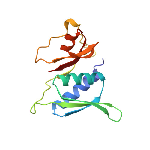
    Prokaryotic ribosomal protein S8 is an important RNA-binding protein that occupies a central position within the small ribosomal subunit. It interacts extensively with 16S rRNA and is crucial for the correct folding of the central domain of the rRNA. S8 also controls the synthesis of several ribosomal proteins by binding to mRNA. It binds specifically to very similar sites in the two RNA molecules. S8 is divided into two tightly associated domains and contains three regions that are proposed to interact with other ribosomal components: two potential RNA-binding sites, and a hydrophobic patch that may interact with a complementary hydrophobic region of S5. The N-terminal domain fold is found in several proteins including two that bind double-stranded DNA. These multiple RNA-binding sites are consistent with the role of S8 in organizing the central domain and agree with the latest models of the 16S RNA which show that the S8 location coincides with a region of complicated nucleic-acid structure. The presence in a wide variety of proteins of a region homologous to the N-terminal domain supports the idea that ribosomal proteins must represent some of the earliest protein molecules.


  • Organizational Affiliation
    • Department of Microbiology, Duke University Medical Center, Durham, NC 27710, USA.

Macromolecule Content 

  • Total Structure Weight: 29.31 kDa 
  • Atom Count: 2,175 
  • Modeled Residue Count: 260 
  • Deposited Residue Count: 260 
  • Unique protein chains: 1

Macromolecules

Find similar proteins by:|  3D Structure
Entity ID: 1
MoleculeChains  Sequence LengthOrganismDetailsImage
RIBOSOMAL PROTEIN S8
A, B
130Geobacillus stearothermophilusMutation(s): 0 
Gene Names: BACILLUS STEAROTHERMOPHILUS
UniProt
Find proteins for P56209 (Geobacillus stearothermophilus)
Explore P56209 
Go to UniProtKB:  P56209
Entity Groups
Sequence Clusters30% Identity50% Identity70% Identity90% Identity95% Identity100% Identity
UniProt GroupP56209
Sequence Annotations
Expand
Reference Sequence

Experimental Data & Validation

Experimental Data

  • Method: X-RAY DIFFRACTION
  • Resolution: 1.90 Å
  • R-Value Free:  0.288 (Depositor) 
  • R-Value Work:  0.222 (Depositor) 
  • R-Value Observed: 0.222 (Depositor) 
Space Group: P 21 21 2
Unit Cell:
Length ( Å )Angle ( ˚ )
a = 80.16α = 90
b = 85.96β = 90
c = 39.59γ = 90
Software Package:
Software NamePurpose
X-PLORmodel building
X-PLORrefinement
DENZOdata reduction
X-PLORphasing

Structure Validation

View Full Validation Report


Entry History 

Deposition Data

Revision History  (Full details and data files)

  • Version 1.0: 1997-03-12
    Type: Initial release
  • Version 1.1: 2008-03-24
    Changes: Version format compliance
  • Version 1.2: 2011-07-13
    Changes: Version format compliance
  • Version 1.3: 2024-02-14
    Changes: Data collection, Database references, Other