1SDN

CRYSTAL STRUCTURE OF A DEACYLATION-DEFECTIVE MUTANT OF PENICILLIN-BINDING PROTEIN 5 MODIFIED BY MERCURY


Experimental Data Snapshot

  • Method: X-RAY DIFFRACTION
  • Resolution: 2.50 Å
  • R-Value Free: 0.241 
  • R-Value Work: 0.171 
  • R-Value Observed: 0.174 

wwPDB Validation   3D Report Full Report


This is version 1.4 of the entry. See complete history


Literature

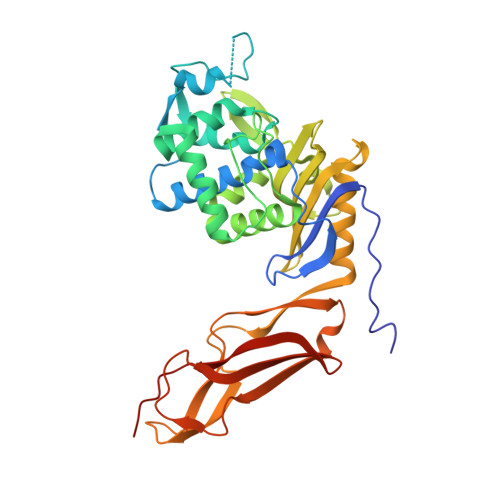
A large displacement of the SXN motif of Cys115-modified penicillin-binding protein 5 from Escherichia coli.

Nicola, G.Fedarovich, A.Nicholas, R.A.Davies, C.

(2005) Biochem J 392: 55-63

  • DOI: https://doi.org/10.1042/BJ20050449
  • Primary Citation of Related Structures:  
    1NZU, 1SDN

  • PubMed Abstract: 

    Penicillin-binding proteins (PBPs), which are the lethal targets of beta-lactam antibiotics, catalyse the final stages of peptidoglycan biosynthesis of the bacterial cell wall. PBP 5 of Escherichia coli is a D-alanine CPase (carboxypeptidase) that has served as a useful model to elucidate the catalytic mechanism of low-molecular-mass PBPs. Previous studies have shown that modification of Cys115 with a variety of reagents results in a loss of CPase activity and a large decrease in the rate of deacylation of the penicilloyl-PBP 5 complex [Tamura, Imae and Strominger (1976) J. Biol. Chem. 251, 414-423; Curtis and Strominger (1978) J. Biol. Chem. 253, 2584-2588]. The crystal structure of wild-type PBP 5 in which Cys115 fortuitously had formed a covalent adduct with 2-mercaptoethanol was solved at 2.0 A (0.2 nm) resolution, and these results provide a structural rationale for how thiol-directed reagents lower the rate of deacylation. When compared with the structure of the unmodified wild-type enzyme, a major change in the architecture of the active site is observed. The two largest differences are the disordering of a loop comprising residues 74-90 and a shift in residues 106-111, which results in the displacement of Ser110 of the SXN active-site motif. These results support the developing hypothesis that the SXN motif of PBP 5, and especially Ser110, is intimately involved in the catalytic mechanism of deacylation.


  • Organizational Affiliation

    Department of Biochemistry and Molecular Biology, Medical University of South Carolina, Charleston, SC 29425, USA.


Macromolecules
Find similar proteins by:  (by identity cutoff)  |  3D Structure
Entity ID: 1
MoleculeChains Sequence LengthOrganismDetailsImage
Penicillin-binding protein 5363Escherichia coliMutation(s): 1 
Gene Names: DACAPFVB0632C0722Z0777ECS0670
EC: 3.4.16.4
UniProt
Find proteins for P0AEB2 (Escherichia coli (strain K12))
Explore P0AEB2 
Go to UniProtKB:  P0AEB2
Entity Groups  
Sequence Clusters30% Identity50% Identity70% Identity90% Identity95% Identity100% Identity
UniProt GroupP0AEB2
Sequence Annotations
Expand
  • Reference Sequence
Small Molecules
Ligands 1 Unique
IDChains Name / Formula / InChI Key2D Diagram3D Interactions
HG
Query on HG

Download Ideal Coordinates CCD File 
B [auth A]MERCURY (II) ION
Hg
BQPIGGFYSBELGY-UHFFFAOYSA-N
Experimental Data & Validation

Experimental Data

  • Method: X-RAY DIFFRACTION
  • Resolution: 2.50 Å
  • R-Value Free: 0.241 
  • R-Value Work: 0.171 
  • R-Value Observed: 0.174 
  • Space Group: P 32
Unit Cell:
Length ( Å )Angle ( ˚ )
a = 50.7α = 90
b = 50.7β = 90
c = 140.3γ = 120
Software Package:
Software NamePurpose
REFMACrefinement
NONIUSdata reduction
SCALEPACKdata scaling
CNSphasing

Structure Validation

View Full Validation Report


Entry History 

Deposition Data

Revision History  (Full details and data files)

  • Version 1.0: 2005-02-22
    Type: Initial release
  • Version 1.1: 2008-04-29
    Changes: Version format compliance
  • Version 1.2: 2011-07-13
    Changes: Version format compliance
  • Version 1.3: 2021-10-27
    Changes: Database references, Derived calculations
  • Version 1.4: 2023-08-23
    Changes: Data collection, Refinement description