1SCJ

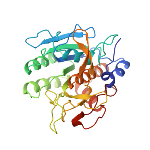
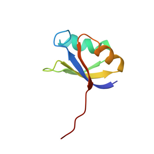
CRYSTAL STRUCTURE OF SUBTILISIN-PROPEPTIDE COMPLEX


Experimental Data Snapshot

  • Method: X-RAY DIFFRACTION
  • Resolution: 2.00 Å
  • R-Value Free: 0.220 
  • R-Value Work: 0.173 
  • R-Value Observed: 0.173 

Starting Models: experimental
View more details

wwPDB Validation   3D Report Full Report


This is version 1.5 of the entry. See complete history


Literature

The crystal structure of an autoprocessed Ser221Cys-subtilisin E-propeptide complex at 2.0 A resolution.

Jain, S.C.Shinde, U.Li, Y.Inouye, M.Berman, H.M.

(1998) J Mol Biol 284: 137-144

  • DOI: https://doi.org/10.1006/jmbi.1998.2161
  • Primary Citation of Related Structures:  
    1SCJ

  • PubMed Abstract: 

    We report here the crystallographic structure determination of an autoprocessed (Ser221Cys)-subtilisin E-propeptide complex at 2.0 A resolution. The subtilisin domain sequence has a single substitution (Ser221Cys) which has been shown to block the maturation process prior to degradation of the propeptide domain (77 residues) that acts as an intramolecular chaperon. This mutation, however, did not prevent the enzyme from cleaving its propeptide domain with a 60-80% efficiency. The current determination is the first example of a subtilisin E-propeptide complex which has been autoprocessed. A previous structure determination of a BPN'-prosegment complex has been reported in which the subtilisin domain was extensively mutated and a calcium binding loop was deleted. Further, in this earlier determination, the complex was formed by the addition of separately expressed propeptide domain. The structure determination reported here provides additional information about the nature of the interaction between the subtilisin and propeptide domains in this complex.


  • Organizational Affiliation

    Department of Chemistry, Rutgers University, 610 Taylor Road, Piscataway, NJ, 08854-8087, USA.


Macromolecules
Find similar proteins by:  (by identity cutoff)  |  3D Structure
Entity ID: 1
MoleculeChains Sequence LengthOrganismDetailsImage
SUBTILISIN E275Bacillus subtilisMutation(s): 1 
EC: 3.4.21.62
UniProt
Find proteins for P04189 (Bacillus subtilis (strain 168))
Explore P04189 
Go to UniProtKB:  P04189
Entity Groups  
Sequence Clusters30% Identity50% Identity70% Identity90% Identity95% Identity100% Identity
UniProt GroupP04189
Sequence Annotations
Expand
  • Reference Sequence
Find similar proteins by:  (by identity cutoff)  |  3D Structure
Entity ID: 2
MoleculeChains Sequence LengthOrganismDetailsImage
SUBTILISIN E71Bacillus subtilisMutation(s): 0 
EC: 3.4.21.62
UniProt
Find proteins for P04189 (Bacillus subtilis (strain 168))
Explore P04189 
Go to UniProtKB:  P04189
Entity Groups  
Sequence Clusters30% Identity50% Identity70% Identity90% Identity95% Identity100% Identity
UniProt GroupP04189
Sequence Annotations
Expand
  • Reference Sequence
Experimental Data & Validation

Experimental Data

  • Method: X-RAY DIFFRACTION
  • Resolution: 2.00 Å
  • R-Value Free: 0.220 
  • R-Value Work: 0.173 
  • R-Value Observed: 0.173 
  • Space Group: P 21 21 2
Unit Cell:
Length ( Å )Angle ( ˚ )
a = 73.544α = 90
b = 92.21β = 90
c = 47.415γ = 90
Software Package:
Software NamePurpose
CNSrefinement
DENZOdata reduction
SCALEPACKdata scaling
CNSphasing

Structure Validation

View Full Validation Report


Entry History 

Deposition Data

Revision History  (Full details and data files)

  • Version 1.0: 1998-12-09
    Type: Initial release
  • Version 1.1: 2008-03-24
    Changes: Version format compliance
  • Version 1.2: 2011-07-13
    Changes: Version format compliance
  • Version 1.3: 2018-03-14
    Changes: Database references
  • Version 1.4: 2023-08-09
    Changes: Database references, Derived calculations, Refinement description
  • Version 1.5: 2024-05-22
    Changes: Data collection