1SBX | pdb_00001sbx

Crystal structure of the Dachshund-homology domain of human SKI


Experimental Data Snapshot

  • Method: X-RAY DIFFRACTION
  • Resolution: 1.65 Å
  • R-Value Free: 
    0.206 (Depositor), 0.212 (DCC) 
  • R-Value Work: 
    0.170 (Depositor), 0.186 (DCC) 
  • R-Value Observed: 
    0.184 (Depositor) 

wwPDB Validation 3D Report Full Report

Validation slider image for 1SBX

This is version 1.3 of the entry. See complete history

Literature

Crystal Structure of the Dachshund Homology Domain of human SKI

Wilson, J.J.Malakhova, M.Zhang, R.Joachimiak, A.Hegde, R.S.

(2004) Structure 12: 785-792

  • DOI: https://doi.org/10.1016/j.str.2004.02.035
  • Primary Citation Related Structures: 
    1SBX

  • PubMed Abstract: 

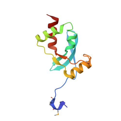
    The nuclear protooncoprotein SKI negatively regulates transforming growth factor-beta (TGF-beta) signaling in cell growth and differentiation. It directly interacts with the Smads and, by various mechanisms, represses the transcription of TGF-beta-responsive genes. SKI is a multidomain protein that includes a domain bearing high sequence similarity with the retinal determination protein Dachshund (the Dachshund homology domain, DHD). The SKI-DHD has been implicated in SMAD-2/3, N-CoR, SKIP, and PML-RARalpha binding. The 1.65 A crystal structure of the Dachshund homology domain of human SKI is reported here. The SKI-DHD adopts a mixed alpha/beta structure which includes features found in the forkhead/winged-helix family of DNA binding proteins, although SKI-DHD is not a DNA binding domain. Residues that form a contiguous surface patch on SKI-DHD are conserved within the Ski/Sno family and with Dachshund, suggesting that this domain may mediate intermolecular interactions common to these proteins.


  • Organizational Affiliation
    • Division of Developmental Biology, Cincinnati Children's Hospital Medical Center and University of Cincinnati College of Medicine, 3333 Burnet Avenue, Cincinnati, OH 45229, USA.

Macromolecule Content 

  • Total Structure Weight: 11.87 kDa 
  • Atom Count: 944 
  • Modeled Residue Count: 106 
  • Deposited Residue Count: 106 
  • Unique protein chains: 1

Macromolecules

Find similar proteins by:|  3D Structure
Entity ID: 1
MoleculeChains  Sequence LengthOrganismDetailsImage
Ski oncogene106Homo sapiensMutation(s): 3 
Gene Names: SKI
UniProt & NIH Common Fund Data Resources
Find proteins for P12755 (Homo sapiens)
Explore P12755 
Go to UniProtKB:  P12755
PHAROS:  P12755
GTEx:  ENSG00000157933 
Entity Groups
Sequence Clusters30% Identity50% Identity70% Identity90% Identity95% Identity100% Identity
UniProt GroupP12755
Sequence Annotations
Expand
Reference Sequence

Small Molecules

Modified Residues  1 Unique
IDChains TypeFormula2D DiagramParent
MSE
Query on MSE
A
L-PEPTIDE LINKINGC5 H11 N O2 SeMET

Experimental Data & Validation

Experimental Data

  • Method: X-RAY DIFFRACTION
  • Resolution: 1.65 Å
  • R-Value Free:  0.206 (Depositor), 0.212 (DCC) 
  • R-Value Work:  0.170 (Depositor), 0.186 (DCC) 
  • R-Value Observed: 0.184 (Depositor) 
Space Group: P 1 21 1
Unit Cell:
Length ( Å )Angle ( ˚ )
a = 25.927α = 90
b = 46.633β = 93.97
c = 35.729γ = 90
Software Package:
Software NamePurpose
CNSrefinement
HKL-2000data reduction
SCALEPACKdata scaling
SOLVEphasing

Structure Validation

View Full Validation Report


Entry History 

Deposition Data

Revision History  (Full details and data files)

  • Version 1.0: 2004-05-25
    Type: Initial release
  • Version 1.1: 2008-04-29
    Changes: Version format compliance
  • Version 1.2: 2011-07-13
    Changes: Version format compliance
  • Version 1.3: 2024-10-30
    Changes: Data collection, Database references, Derived calculations, Structure summary