1SB7 | pdb_00001sb7

Crystal structure of the E.coli pseudouridine synthase TruD


Experimental Data Snapshot

  • Method: X-RAY DIFFRACTION
  • Resolution: 2.20 Å
  • R-Value Free: 
    0.254 (Depositor), 0.259 (DCC) 
  • R-Value Work: 
    0.216 (Depositor), 0.220 (DCC) 
  • R-Value Observed: 
    0.216 (Depositor) 

wwPDB Validation 3D Report Full Report

Validation slider image for 1SB7

This is version 1.3 of the entry. See complete history

Literature

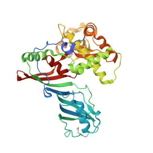
Crystal structure of the highly divergent pseudouridine synthase TruD reveals a circular permutation of a conserved fold.

Hoang, C.Ferre-D'Amare, A.R.

(2004) RNA 10: 1026-1033

  • DOI: https://doi.org/10.1261/rna.7240504
  • Primary Citation Related Structures: 
    1SB7

  • PubMed Abstract: 

    The pseudouridine (Psi) synthases Pus7p and TruD define a family of RNA-modifying enzymes with no sequence similarity to previously characterized Psi synthases. The 2.2 A resolution structure of Escherichia coli TruD reveals a U-shaped molecule with a catalytic domain that superimposes closely on that of other Psi synthases. A domain that appears to be unique to TruD/Pus7p family enzymes hinges over the catalytic domain, possibly serving to clasp the substrate RNAs. The active site comprises residues that are conserved in other Psi synthases, although at least one comes from a structurally distinct part of the protein. Remarkably, the connectivity of the structural elements of the TruD catalytic domain is a circular permutation of that of its paralogs. Because the sequence of the permuted segment, a beta-strand that bisects the catalytic domain, is conserved among orthologs from bacteria, archaea and eukarya, the permutation likely happened early in evolution.


  • Organizational Affiliation
    • Division of Basic Sciences, Fred Hutchinson Cancer Research Center, 1100 Fairview Avenue North, Seattle, WA 98109-1024, USA.

Macromolecule Content 

  • Total Structure Weight: 83.38 kDa 
  • Atom Count: 5,723 
  • Modeled Residue Count: 686 
  • Deposited Residue Count: 736 
  • Unique protein chains: 1

Macromolecules

Find similar proteins by:|  3D Structure
Entity ID: 1
MoleculeChains  Sequence LengthOrganismDetailsImage
tRNA pseudouridine synthase D
A, B
368Escherichia coliMutation(s): 4 
Gene Names: TRUDB2745
EC: 4.2.1.70 (PDB Primary Data), 5.4.99.27 (UniProt)
UniProt
Find proteins for Q57261 (Escherichia coli (strain K12))
Explore Q57261 
Go to UniProtKB:  Q57261
Entity Groups
Sequence Clusters30% Identity50% Identity70% Identity90% Identity95% Identity100% Identity
UniProt GroupQ57261
Sequence Annotations
Expand
Reference Sequence

Small Molecules

Modified Residues  1 Unique
IDChains TypeFormula2D DiagramParent
MSE
Query on MSE
A, B
L-PEPTIDE LINKINGC5 H11 N O2 SeMET

Experimental Data & Validation

Experimental Data

  • Method: X-RAY DIFFRACTION
  • Resolution: 2.20 Å
  • R-Value Free:  0.254 (Depositor), 0.259 (DCC) 
  • R-Value Work:  0.216 (Depositor), 0.220 (DCC) 
  • R-Value Observed: 0.216 (Depositor) 
Space Group: P 21 21 21
Unit Cell:
Length ( Å )Angle ( ˚ )
a = 63.665α = 90
b = 108.78β = 90
c = 111.855γ = 90
Software Package:
Software NamePurpose
CNSrefinement
DENZOdata reduction
SCALEPACKdata scaling
SOLVEphasing

Structure Validation

View Full Validation Report


Entry History 

Deposition Data

Revision History  (Full details and data files)

  • Version 1.0: 2004-06-29
    Type: Initial release
  • Version 1.1: 2008-04-29
    Changes: Version format compliance
  • Version 1.2: 2011-07-13
    Changes: Non-polymer description, Version format compliance
  • Version 1.3: 2024-10-30
    Changes: Data collection, Database references, Derived calculations, Structure summary