1S97 | pdb_00001s97

DPO4 with GT mismatch


Experimental Data Snapshot

  • Method: X-RAY DIFFRACTION
  • Resolution: 2.40 Å
  • R-Value Free: 
    0.269 (Depositor), 0.257 (DCC) 
  • R-Value Work: 
    0.218 (Depositor), 0.207 (DCC) 
  • R-Value Observed: 
    0.238 (Depositor) 

Starting Model: experimental
View more details

wwPDB Validation 3D Report Full Report

Validation slider image for 1S97

Ligand Structure Quality Assessment 


This is version 1.3 of the entry. See complete history

Literature

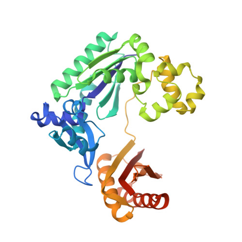
Dpo4 is hindered in extending a G.T mismatch by a reverse wobble

Trincao, J.Johnson, R.E.Wolfle, W.T.Escalante, C.R.Prakash, S.Prakash, L.Aggarwal, A.K.

(2004) Nat Struct Mol Biol 11: 457-462

  • DOI: https://doi.org/10.1038/nsmb755
  • Primary Citation Related Structures: 
    1S97, 1S9F

  • PubMed Abstract: 

    The ability or inability of a DNA polymerase to extend a mispair directly affects the establishment of genomic mutations. We report here kinetic analyses of the ability of Dpo4, a Y-family polymerase from Sulfolobus solfataricus, to extend from all mispairs opposite a template G or T. Dpo4 is equally inefficient at extending these mispairs, which include, surprisingly, a G.T mispair expected to conform closely to Watson-Crick geometry. To elucidate the basis of this, we solved the structure of Dpo4 bound to G.T-mispaired primer template in the presence of an incoming nucleotide. As a control, we also determined the structure of Dpo4 bound to a matched A-T base pair at the primer terminus. The structures offer a basis for the low efficiency of Dpo4 in extending a G.T mispair: a reverse wobble that deflects the primer 3'-OH away from the incoming nucleotide.


  • Organizational Affiliation
    • Structural Biology Program, Department of Physiology and Biophysics, Mount Sinai School of Medicine, Box 1677, 1425 Madison Avenue, New York, New York 10029, USA.

Macromolecule Content 

  • Total Structure Weight: 201 kDa 
  • Atom Count: 14,001 
  • Modeled Residue Count: 1,484 
  • Deposited Residue Count: 1,532 
  • Unique protein chains: 1
  • Unique nucleic acid chains: 2

Macromolecules


Find similar proteins by:|  3D Structure
Entity ID: 3
MoleculeChains  Sequence LengthOrganismDetailsImage
DNA polymerase IVI [auth A],
J [auth B],
K [auth C],
L [auth D]
352Saccharolobus solfataricusMutation(s): 0 
Gene Names: DBHDPO4SSO2448
EC: 2.7.7.7
UniProt
Find proteins for Q97W02 (Saccharolobus solfataricus (strain ATCC 35092 / DSM 1617 / JCM 11322 / P2))
Explore Q97W02 
Go to UniProtKB:  Q97W02
Entity Groups
Sequence Clusters30% Identity50% Identity70% Identity90% Identity95% Identity100% Identity
UniProt GroupQ97W02
Sequence Annotations
Expand
Reference Sequence
Find similar nucleic acids by:  Sequence
Entity ID: 1
MoleculeChains LengthOrganismImage
5'-D(*GP*GP*GP*GP*GP*AP*AP*GP*GP*AP*CP*TP*G)-3'A [auth E],
C [auth F],
E [auth G],
G [auth H]
13N/A
Sequence Annotations
Expand
Reference Sequence
Find similar nucleic acids by:  Sequence
Entity ID: 2
MoleculeChains LengthOrganismImage
5'-D(*T*TP*CP*AP*GP*TP*AP*GP*TP*CP*CP*TP*TP*CP*CP*CP*CP*C)-3'B [auth I],
D [auth J],
F [auth K],
H [auth L]
18N/A
Sequence Annotations
Expand
Reference Sequence

Small Molecules

Ligands 2 Unique
IDChains Name / Formula / InChI Key2D Diagram3D Interactions
DCT

Query on DCT


Download:Ideal Coordinates CCD File
N [auth A],
P [auth B],
R [auth C],
T [auth D]
2',3'-DIDEOXYCYTIDINE 5'-TRIPHOSPHATE
C9 H16 N3 O12 P3
ARLKCWCREKRROD-POYBYMJQSA-N
CA

Query on CA


Download:Ideal Coordinates CCD File
M [auth A],
O [auth B],
Q [auth C],
S [auth D]
CALCIUM ION
Ca
BHPQYMZQTOCNFJ-UHFFFAOYSA-N

Experimental Data & Validation

Experimental Data

  • Method: X-RAY DIFFRACTION
  • Resolution: 2.40 Å
  • R-Value Free:  0.269 (Depositor), 0.257 (DCC) 
  • R-Value Work:  0.218 (Depositor), 0.207 (DCC) 
  • R-Value Observed: 0.238 (Depositor) 
Space Group: P 1 21 1
Unit Cell:
Length ( Å )Angle ( ˚ )
a = 104.433α = 90
b = 100.644β = 94.89
c = 110.426γ = 90
Software Package:
Software NamePurpose
CNSrefinement
HKL-2000data reduction
SCALEPACKdata scaling
CNSphasing

Structure Validation

View Full Validation Report


Ligand Structure Quality Assessment 


Entry History 

Deposition Data

Revision History  (Full details and data files)

  • Version 1.0: 2004-04-27
    Type: Initial release
  • Version 1.1: 2008-04-29
    Changes: Version format compliance
  • Version 1.2: 2011-07-13
    Changes: Version format compliance
  • Version 1.3: 2023-08-23
    Changes: Data collection, Database references, Derived calculations, Refinement description