NMR structure determination of a modified DNA oligonucleotide containing a new intercalating nucleic acid.
Nielsen, C.B., Petersen, M., Pedersen, E.B., Hansen, P.E., Christensen, U.B.(2004) Bioconjug Chem 15: 260-269
- PubMed: 15025521 Search on PubMed
- DOI: https://doi.org/10.1021/bc0341932
- Primary Citation Related Structures:
1S88 - PubMed Abstract:
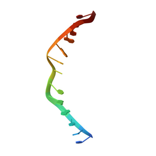
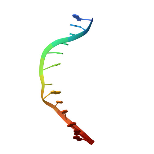
The intercalating nucleic acid (INA) presented in this paper is a novel 1-O-(1-pyrenylmethyl)glycerol DNA intercalator that induces high thermal affinity for complementary DNA. The duplex examined contained two INA intercalators, denoted X, inserted directly opposite each other: d(C(1)T(2)C(3)A(4)A(5)C(6)X(7)C(8)A(9)A(10)G(11)C(12)T(13)):d(A(14)G(15)C(16)T(17)-T(18)G(19)X(20)G(21)T(22)T(23)G(24)A(25)G(26)). Unlike most other nucleotide analogues, DNA with INA inserted has a lower affinity for hybridizing to complementary DNA with an INA inserted directly opposite than to complementary unmodified DNA. In this study we used two-dimensional (1)H NMR spectroscopy to determine a high-resolution solution structure of the weak INA-INA duplex. A modified ISPA approach was used to obtain interproton distance bounds from NOESY cross-peak intensities. These distance bounds were used as restraints in molecular dynamics (rMD) calculations. Twenty final structures were generated for the duplex from a B-type DNA starting structure. The root-mean-square deviation (RMSD) of the coordinates for the 20 structures of the complex was 1.95 A. This rather large value, together with broad lines in the area of insertion, reflect the high degree of internal motion in the complex. The determination of the structure revealed that both intercalators were situated in the center of the helix, stacking with each other and the neighboring nucleobases. The intercalation of the INAs caused an unwinding of the helix in the insertion area, creating a ladderlike structure. The structural changes observed upon intercalation were mainly of local character; however, a broadening of the minor groove was found throughout the helix.
- Nucleic Acid Center, Department of Chemistry, University of Southern Denmark, DK-5230 Odense M, Denmark. cbn@chem.sdu.dk
Organizational Affiliation: