1S6A

The X-ray structure of the cyanobacteria Synechocystis hemoglobin "cyanoglobin" with azide ligand


Experimental Data Snapshot

  • Method: X-RAY DIFFRACTION
  • Resolution: 1.69 Å
  • R-Value Free: 0.219 
  • R-Value Work: 0.197 
  • R-Value Observed: 0.198 

wwPDB Validation   3D Report Full Report


Ligand Structure Quality Assessment 


This is version 1.3 of the entry. See complete history


Literature

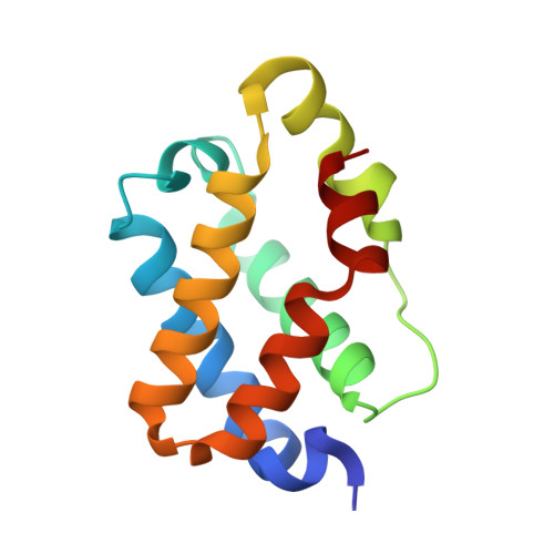
Crystallographic analysis of synechocystis cyanoglobin reveals the structural changes accompanying ligand binding in a hexacoordinate hemoglobin.

Trent III, J.T.Kundu, S.Hoy, J.A.Hargrove, M.S.

(2004) J Mol Biol 341: 1097-1108

  • DOI: https://doi.org/10.1016/j.jmb.2004.05.070
  • Primary Citation of Related Structures:  
    1S69, 1S6A

  • PubMed Abstract: 

    The crystal structures of cyanide and azide-bound forms of the truncated hemoglobin from Synechocystis are presented at 1.8 angstroms resolution. A comparison with the structure of the endogenously liganded protein reveals a conformational shift unprecedented in hemoglobins, and provides the first picture of a hexacoordinate hemoglobin in both the bis-histidyl and the exogenously coordinated states. The structural changes between the different conformations are confined to two regions of the protein; the B helix, and the E helix, including the EF loop. A molecular "hinge" controlling movement of the E helix is observed in the EF loop, which is composed of three principal structural elements: Arg64, the heme-d-propionate, and a three-residue extension of the F helix. Additional features of the structural transition between the two protein conformations are discussed as they relate to the complex ligand-binding behavior observed in hexacoordinate hemoglobins, and the potential physiological function of this class of proteins.


  • Organizational Affiliation

    Department of Biochemistry, Biophysics and Molecular Biology, Iowa State University, Ames, IA 50010, USA.


Macromolecules
Find similar proteins by:  (by identity cutoff)  |  3D Structure
Entity ID: 1
MoleculeChains Sequence LengthOrganismDetailsImage
Cyanoglobin124Synechocystis sp. PCC 6803Mutation(s): 0 
Gene Names: GLBNSLR2097
UniProt
Find proteins for P73925 (Synechocystis sp. (strain PCC 6803 / Kazusa))
Explore P73925 
Go to UniProtKB:  P73925
Entity Groups  
Sequence Clusters30% Identity50% Identity70% Identity90% Identity95% Identity100% Identity
UniProt GroupP73925
Sequence Annotations
Expand
  • Reference Sequence
Experimental Data & Validation

Experimental Data

  • Method: X-RAY DIFFRACTION
  • Resolution: 1.69 Å
  • R-Value Free: 0.219 
  • R-Value Work: 0.197 
  • R-Value Observed: 0.198 
  • Space Group: I 41
Unit Cell:
Length ( Å )Angle ( ˚ )
a = 83.35α = 90
b = 83.35β = 90
c = 65.18γ = 90
Software Package:
Software NamePurpose
REFMACrefinement
CRYSTALdata reduction
d*TREKdata scaling
AMoREphasing

Structure Validation

View Full Validation Report


Ligand Structure Quality Assessment 


Entry History 

Deposition Data

Revision History  (Full details and data files)

  • Version 1.0: 2004-09-21
    Type: Initial release
  • Version 1.1: 2008-04-29
    Changes: Version format compliance
  • Version 1.2: 2011-07-13
    Changes: Source and taxonomy, Version format compliance
  • Version 1.3: 2023-08-23
    Changes: Data collection, Database references, Derived calculations, Refinement description