1S5Z | pdb_00001s5z

NDP kinase in complex with adenosine phosphonoacetic acid


Experimental Data Snapshot

  • Method: X-RAY DIFFRACTION
  • Resolution: 2.00 Å
  • R-Value Free: 
    0.239 (Depositor), 0.240 (DCC) 
  • R-Value Work: 
    0.187 (Depositor), 0.187 (DCC) 

Starting Model: experimental
View more details

wwPDB Validation 3D Report Full Report

Validation slider image for 1S5Z

Ligand Structure Quality Assessment 


This is version 1.3 of the entry. See complete history

Literature

Adenosine phosphonoacetic acid is slowly metabolized by NDP kinase.

Chen, Y.Morera, S.Pasti, C.Angusti, A.Solaroli, N.Veron, M.Janin, J.Manfredini, S.Deville-Bonne, D.

(2005) Med Chem 1: 529-536

  • DOI: https://doi.org/10.2174/157340605774598162
  • Primary Citation Related Structures: 
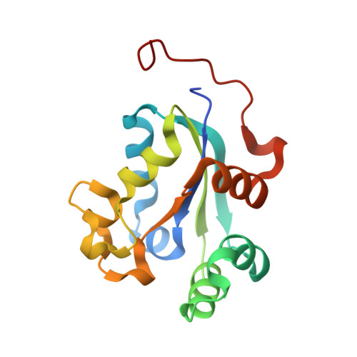
    1S5Z

  • PubMed Abstract: 

    NDP kinase catalyzes the last step in the phosphorylation of nucleotides. It is also involved in the activation by cellular kinases of nucleoside analogs used in antiviral therapies. Adenosine phosphonoacetic acid, a close analog of ADP already proposed as an inhibitor of ribonucleotide reductase, was found to be a poor substrate for human NDP kinase, as well as a weak inhibitor with an equilibrium dissociation constant of 0.6 mM to be compared to 0.025 mM for ADP. The X-ray structure of a complex of adenosine phosphonoacetic acid and the NDP kinase from Dictyostelium was determined to 2.0 A resolution showing that the analog adopts a binding mode similar to ADP, but that no magnesium ion is present at the active site. As ACP may also interfere with other cellular kinases, its potential as a drug targeting NDP kinase or ribonucleotide reductase is likely to be limited due to strong side effects. The design of new molecules with a narrower specificity and a stronger affinity will benefit from the detailed knowledge of the complex ACP-NDP kinase.


  • Organizational Affiliation
    • Laboratoire d'Enzymologie et de Biochimie Structurales, CNRS UPR 9063, Paris, France.

Macromolecule Content 

  • Total Structure Weight: 101.38 kDa 
  • Atom Count: 8,058 
  • Modeled Residue Count: 900 
  • Deposited Residue Count: 930 
  • Unique protein chains: 1

Macromolecules

Find similar proteins by:|  3D Structure
Entity ID: 1
MoleculeChains  Sequence LengthOrganismDetailsImage
Nucleoside diphosphate kinase, cytosolic
A, B, C, D, E
A, B, C, D, E, F
155Dictyostelium discoideumMutation(s): 0 
Gene Names: NDKCNDKBGIP17DD_02290DD_02289
EC: 2.7.4.6
UniProt
Find proteins for P22887 (Dictyostelium discoideum)
Explore P22887 
Go to UniProtKB:  P22887
Entity Groups
Sequence Clusters30% Identity50% Identity70% Identity90% Identity95% Identity100% Identity
UniProt GroupP22887
Sequence Annotations
Expand
Reference Sequence

Small Molecules

Experimental Data & Validation

Experimental Data

  • Method: X-RAY DIFFRACTION
  • Resolution: 2.00 Å
  • R-Value Free:  0.239 (Depositor), 0.240 (DCC) 
  • R-Value Work:  0.187 (Depositor), 0.187 (DCC) 
Space Group: P 1 21 1
Unit Cell:
Length ( Å )Angle ( ˚ )
a = 69.38α = 90
b = 104.594β = 118.28
c = 69.569γ = 90
Software Package:
Software NamePurpose
DENZOdata reduction
SCALEPACKdata scaling
AMoREphasing
CNSrefinement

Structure Validation

View Full Validation Report


Ligand Structure Quality Assessment 


Entry History 

Deposition Data

Revision History  (Full details and data files)

  • Version 1.0: 2005-02-01
    Type: Initial release
  • Version 1.1: 2008-04-29
    Changes: Version format compliance
  • Version 1.2: 2011-07-13
    Changes: Version format compliance
  • Version 1.3: 2023-08-23
    Changes: Data collection, Database references, Derived calculations, Refinement description