1S57

crystal structure of nucleoside diphosphate kinase 2 from Arabidopsis


Experimental Data Snapshot

  • Method: X-RAY DIFFRACTION
  • Resolution: 1.80 Å
  • R-Value Free: 0.241 
  • R-Value Work: 0.212 
  • R-Value Observed: 0.212 

wwPDB Validation   3D Report Full Report


This is version 1.3 of the entry. See complete history


Literature

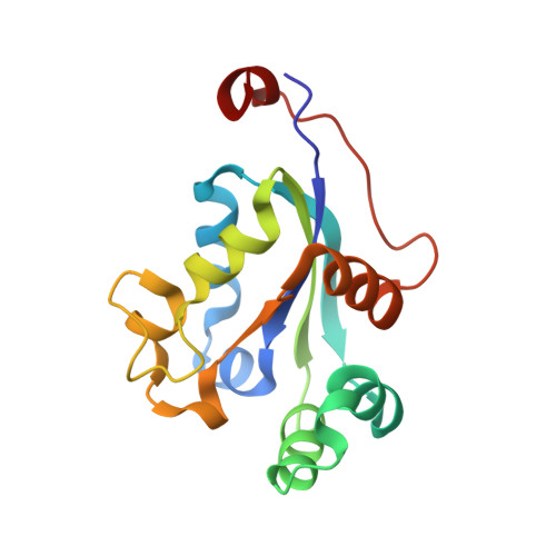
Structural analysis of Arabidopsis thaliana nucleoside diphosphate kinase-2 for phytochrome-mediated light signaling

Im, Y.J.Kim, J.-I.Shen, Y.Na, Y.Han, Y.-J.Kim, S.-H.Song, P.-S.Eom, S.H.

(2004) J Mol Biol 343: 659-670

  • DOI: https://doi.org/10.1016/j.jmb.2004.08.054
  • Primary Citation of Related Structures:  
    1S57, 1S59, 1U8W

  • PubMed Abstract: 

    In plants, nucleoside diphosphate kinases (NDPKs) play a key role in the signaling of both stress and light. However, little is known about the structural elements involved in their function. Of the three NDPKs (NDPK1-NDPK3) expressed in Arabidopsis thaliana, NDPK2 is involved in phytochrome-mediated signal transduction. In this study, we found that the binding of dNDP or NTP to NDPK2 strengthens the interaction significantly between activated phytochrome and NDPK2. To better understand the structural basis of the phytochrome-NDPK2 interaction, we determined the X-ray structures of NDPK1, NDPK2, and dGTP-bound NDPK2 from A.thaliana at 1.8A, 2.6A, and 2.4A, respectively. The structures showed that nucleotide binding caused a slight conformational change in NDPK2 that was confined to helices alphaA and alpha2. This suggests that the presence of nucleotide in the active site and/or the evoked conformational change contributes to the recognition of NDPK2 by activated phytochrome. In vitro binding assays showed that only NDPK2 interacted specifically with the phytochrome and the C-terminal regulatory domain of phytochrome is involved in the interaction. A domain swap experiment between NDPK1 and NDPK2 showed that the variable C-terminal region of NDPK2 is important for the activation by phytochrome. The structure of Arabidopsis NDPK1 and NDPK2 showed that the isoforms share common electrostatic surfaces at the nucleotide-binding site, but the variable C-terminal regions have distinct electrostatic charge distributions. These findings suggest that the binding of nucleotide to NDPK2 plays a regulatory role in phytochrome signaling and that the C-terminal extension of NDPK2 provides a potential binding surface for the specific interaction with phytochromes.


  • Organizational Affiliation

    Department of Life Science, Gwangju Institute of Science and Technology, Gwangju 500-712, South Korea.


Macromolecules
Find similar proteins by:  (by identity cutoff)  |  3D Structure
Entity ID: 1
MoleculeChains Sequence LengthOrganismDetailsImage
Nucleoside diphosphate kinase II
A, B, C, D, E
A, B, C, D, E, F
153Arabidopsis thalianaMutation(s): 0 
Gene Names: NDPK2
EC: 2.7.4.6
UniProt
Find proteins for O64903 (Arabidopsis thaliana)
Explore O64903 
Go to UniProtKB:  O64903
Entity Groups  
Sequence Clusters30% Identity50% Identity70% Identity90% Identity95% Identity100% Identity
UniProt GroupO64903
Sequence Annotations
Expand
  • Reference Sequence
Small Molecules
Ligands 2 Unique
IDChains Name / Formula / InChI Key2D Diagram3D Interactions
EPE
Query on EPE

Download Ideal Coordinates CCD File 
DA [auth F]
J [auth A]
N [auth B]
R [auth C]
S [auth C]
DA [auth F],
J [auth A],
N [auth B],
R [auth C],
S [auth C],
W [auth D]
4-(2-HYDROXYETHYL)-1-PIPERAZINE ETHANESULFONIC ACID
C8 H18 N2 O4 S
JKMHFZQWWAIEOD-UHFFFAOYSA-N
SO4
Query on SO4

Download Ideal Coordinates CCD File 
AA [auth F]
BA [auth F]
CA [auth F]
G [auth A]
H [auth A]
AA [auth F],
BA [auth F],
CA [auth F],
G [auth A],
H [auth A],
I [auth A],
K [auth B],
L [auth B],
M [auth B],
O [auth C],
P [auth C],
Q [auth C],
T [auth D],
U [auth D],
V [auth D],
X [auth E],
Y [auth E],
Z [auth E]
SULFATE ION
O4 S
QAOWNCQODCNURD-UHFFFAOYSA-L
Experimental Data & Validation

Experimental Data

  • Method: X-RAY DIFFRACTION
  • Resolution: 1.80 Å
  • R-Value Free: 0.241 
  • R-Value Work: 0.212 
  • R-Value Observed: 0.212 
  • Space Group: P 21 21 21
Unit Cell:
Length ( Å )Angle ( ˚ )
a = 69.394α = 90
b = 108.855β = 90
c = 119.324γ = 90
Software Package:
Software NamePurpose
CNSrefinement
HKL-2000data reduction
SCALEPACKdata scaling
AMoREphasing

Structure Validation

View Full Validation Report


Entry History 

Deposition Data

Revision History  (Full details and data files)

  • Version 1.0: 2004-11-30
    Type: Initial release
  • Version 1.1: 2008-04-29
    Changes: Version format compliance
  • Version 1.2: 2011-07-13
    Changes: Version format compliance
  • Version 1.3: 2023-10-25
    Changes: Data collection, Database references, Derived calculations, Refinement description