1RY6 | pdb_00001ry6

Crystal Structure of Internal Kinesin Motor Domain


Experimental Data Snapshot

  • Method: X-RAY DIFFRACTION
  • Resolution: 1.60 Å
  • R-Value Free: 
    0.231 (Depositor), 0.214 (DCC) 
  • R-Value Work: 
    0.205 (Depositor), 0.217 (DCC) 
  • R-Value Observed: 
    0.207 (Depositor) 

Starting Model: experimental
View more details

wwPDB Validation 3D Report Full Report

Validation slider image for 1RY6

This is version 1.4 of the entry. See complete history

Literature

Structure of a kinesin microtubule depolymerization machine.

Shipley, K.Hekmat-Nejad, M.Turner, J.Moores, C.Anderson, R.Milligan, R.Sakowicz, R.Fletterick, R.

(2004) EMBO J 23: 1422-1432

  • DOI: https://doi.org/10.1038/sj.emboj.7600165
  • Primary Citation Related Structures: 
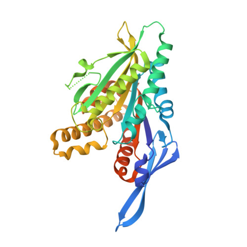
    1RY6

  • PubMed Abstract: 

    With their ability to depolymerize microtubules (MTs), KinI kinesins are the rogue members of the kinesin family. Here we present the 1.6 A crystal structure of a KinI motor core from Plasmodium falciparum, which is sufficient for depolymerization in vitro. Unlike all published kinesin structures to date, nucleotide is not present, and there are noticeable differences in loop regions L6 and L10 (the plus-end tip), L2 and L8 and in switch II (L11 and helix4); otherwise, the pKinI structure is very similar to previous kinesin structures. KinI-conserved amino acids were mutated to alanine, and studied for their effects on depolymerization and ATP hydrolysis. Notably, mutation of three residues in L2 appears to primarily affect depolymerization, rather than general MT binding or ATP hydrolysis. The results of this study confirm the suspected importance of loop 2 for KinI function, and provide evidence that KinI is specialized to hydrolyze ATP after initiating depolymerization.


  • Organizational Affiliation
    • Graduate Group in Biophysics, University of California, San Francisco, CA, USA.

Macromolecule Content 

  • Total Structure Weight: 40.86 kDa 
  • Atom Count: 2,829 
  • Modeled Residue Count: 319 
  • Deposited Residue Count: 360 
  • Unique protein chains: 1

Macromolecules

Find similar proteins by:|  3D Structure
Entity ID: 1
MoleculeChains  Sequence LengthOrganismDetailsImage
INTERNAL KINESIN360Plasmodium falciparumMutation(s): 0 
Gene Names: PFL2165W
UniProt
Find proteins for Q8I4Y0 (Plasmodium falciparum (isolate 3D7))
Explore Q8I4Y0 
Go to UniProtKB:  Q8I4Y0
Entity Groups
Sequence Clusters30% Identity50% Identity70% Identity90% Identity95% Identity100% Identity
UniProt GroupQ8I4Y0
Sequence Annotations
Expand
Reference Sequence

Small Molecules

Ligands 1 Unique
IDChains Name / Formula / InChI Key2D Diagram3D Interactions
SO4

Query on SO4


Download:Ideal Coordinates CCD File
B [auth A]SULFATE ION
O4 S
QAOWNCQODCNURD-UHFFFAOYSA-L

Experimental Data & Validation

Experimental Data

  • Method: X-RAY DIFFRACTION
  • Resolution: 1.60 Å
  • R-Value Free:  0.231 (Depositor), 0.214 (DCC) 
  • R-Value Work:  0.205 (Depositor), 0.217 (DCC) 
  • R-Value Observed: 0.207 (Depositor) 
Space Group: P 32 2 1
Unit Cell:
Length ( Å )Angle ( ˚ )
a = 105.588α = 90
b = 105.588β = 90
c = 84.771γ = 120
Software Package:
Software NamePurpose
REFMACrefinement
DENZOdata reduction
SCALEPACKdata scaling
CNSphasing

Structure Validation

View Full Validation Report


Entry History 

Deposition Data

Revision History  (Full details and data files)

  • Version 1.0: 2004-04-13
    Type: Initial release
  • Version 1.1: 2008-04-29
    Changes: Version format compliance
  • Version 1.2: 2011-07-13
    Changes: Version format compliance
  • Version 1.3: 2022-12-21
    Changes: Database references, Derived calculations
  • Version 1.4: 2023-09-20
    Changes: Data collection, Refinement description