1RXE | pdb_00001rxe

ArsC complexed with MNB


Experimental Data Snapshot

  • Method: X-RAY DIFFRACTION
  • Resolution: 1.70 Å
  • R-Value Free: 
    0.212 (Depositor), 0.216 (DCC) 
  • R-Value Work: 
    0.195 (Depositor), 0.198 (DCC) 
  • R-Value Observed: 
    0.196 (Depositor) 

wwPDB Validation 3D Report Full Report

Validation slider image for 1RXE

Ligand Structure Quality Assessment 


This is version 1.4 of the entry. See complete history

Literature

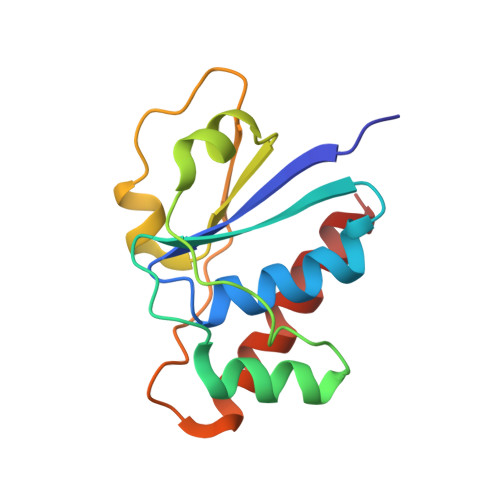
The structure of a triple mutant of pI258 arsenate reductase from Staphylococcus aureus and its 5-thio-2-nitrobenzoic acid adduct.

Messens, J.Van Molle, I.Vanhaesebrouck, P.Van Belle, K.Wahni, K.Martins, J.C.Wyns, L.Loris, R.

(2004) Acta Crystallogr D Biol Crystallogr 60: 1180-1184

  • DOI: https://doi.org/10.1107/S0907444904007334
  • Primary Citation Related Structures: 
    1RXE, 1RXI

  • PubMed Abstract: 

    Structural insights into formation of the complex between the ubiquitous thiol-disulfide oxidoreductase thioredoxin and its oxidized substrate are under-documented owing to its entropical instability. In vitro, it is possible via a reaction with 5,5'-dithiobis-(2-nitrobenzoic acid) to make a stable mixed-disulfide complex between thioredoxin from Staphylococcus aureus and one of its substrates, oxidized pI258 arsenate reductase (ArsC) from S. aureus. In the absence of the crystal structure of an ArsC-thioredoxin complex, the structures of two precursors of the complex, the ArsC triple mutant ArsC C10SC15AC82S and its 5-thio-2-nitrobenzoic acid (TNB) adduct, were determined. The ArsC triple mutant has a structure very similar to that of the reduced form of wild-type ArsC, with a folded redox helix and a buried catalytic Cys89. In the adduct form, the TNB molecule is buried in a hydrophobic pocket and the disulfide bridge between TNB and Cys89 is sterically inaccessible to thioredoxin. In order to form a mixed disulfide between ArsC and thioredoxin, a change in the orientation of the TNB-Cys89 disulfide in the structure is necessary.


  • Organizational Affiliation
    • Laboratorium voor Ultrastructuur, Vlaams Interuniversitair Instituut voor Biotechnologie (VIB), Vrije Universiteit Brussel, Pleinlaan 2, B-1050 Brussel, Belgium. joris.messens@vub.ac.be

Macromolecule Content 

  • Total Structure Weight: 15.11 kDa 
  • Atom Count: 1,166 
  • Modeled Residue Count: 131 
  • Deposited Residue Count: 131 
  • Unique protein chains: 1

Macromolecules

Find similar proteins by:|  3D Structure
Entity ID: 1
MoleculeChains  Sequence LengthOrganismDetailsImage
Arsenate reductase131Staphylococcus aureusMutation(s): 3 
Gene Names: ARSCSAP018
EC: 1.20.4.1 (PDB Primary Data), 1.20.4.4 (UniProt)
UniProt
Find proteins for P0A006 (Staphylococcus aureus)
Explore P0A006 
Go to UniProtKB:  P0A006
Entity Groups
Sequence Clusters30% Identity50% Identity70% Identity90% Identity95% Identity100% Identity
UniProt GroupP0A006
Sequence Annotations
Expand
Reference Sequence

Experimental Data & Validation

Experimental Data

  • Method: X-RAY DIFFRACTION
  • Resolution: 1.70 Å
  • R-Value Free:  0.212 (Depositor), 0.216 (DCC) 
  • R-Value Work:  0.195 (Depositor), 0.198 (DCC) 
  • R-Value Observed: 0.196 (Depositor) 
Space Group: P 21 21 21
Unit Cell:
Length ( Å )Angle ( ˚ )
a = 33.146α = 90
b = 34.377β = 90
c = 102.325γ = 90
Software Package:
Software NamePurpose
DENZOdata reduction
SCALEPACKdata scaling
AMoREphasing
CNSrefinement

Structure Validation

View Full Validation Report


Ligand Structure Quality Assessment 


Entry History 

Deposition Data

Revision History  (Full details and data files)

  • Version 1.0: 2004-06-01
    Type: Initial release
  • Version 1.1: 2008-04-29
    Changes: Version format compliance
  • Version 1.2: 2011-07-13
    Changes: Version format compliance
  • Version 1.3: 2021-10-27
    Changes: Data collection, Database references, Derived calculations
  • Version 1.4: 2024-10-30
    Changes: Data collection, Structure summary