Electrostatic modification of the active site of myoglobin: characterization of the proximal Ser92Asp variant.
Lloyd, E., Burk, D.L., Ferrer, J.C., Maurus, R., Doran, J., Carey, P.R., Brayer, G.D., Mauk, A.G.(1996) Biochemistry 35: 11901-11912
- PubMed: 8794773 Search on PubMed
- DOI: https://doi.org/10.1021/bi9608976
- Primary Citation Related Structures:
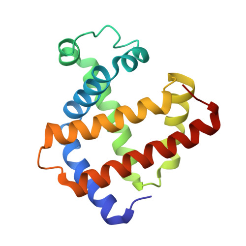
1RSE - PubMed Abstract:
The structural and functional consequences of the introduction of a negatively charged amino acid into the active site of horse heart myoglobin have been investigated by replacement of the proximal Ser92 residue (F7) with an aspartyl residue (Ser92Asp). UV-visible absorption maxima of various ferrous and ferric derivatives and low-temperature EPR spectra of the metaquo (metMb) derivative indicate that the active site coordination geometry has not been perturbed significantly in the variant. 1H-NMR spectroscopy provides direct evidence for the existence of a distal water molecule as the sixth ligand in the oxidized form of the variant at pD 5.7. Spectrophotometric pH titration of the Ser92Asp variant is consistent with this finding and with a pKa = 8.90 +/- 0.02 [25.0 degrees C, mu = 0.10 M (NaCl)] for titration of the distal water molecule, identical to the value reported for the wild-type protein. X-ray crystallography of the metMb derivative indicates that the heme substituents conserve their orientations in the variant protein, except for a slight reorientation of the pyrrole A propionate group to which Ser92 normally hydrogen bonds and reorientation of the carboxyl end of the pyrrole D propionate group. No change is observed in conformation of the proximal (His93) or distal (Wat156) heme ligands. 1H-NMR spectroscopy of the metMbCN form of the protein indicates that a slight rotation of the proximal His93 ligand has occurred in this derivative. Resonance Raman experiments indicate increased conformational heterogeneity in the proximal pocket of the variant. Failure to detect electron density for the Asp residue in the X-ray diffraction map of the variant protein and high average thermal factors for the pyrrole A propionate substituent are consistent with this observation. The variant exhibits novel pH-dependent behavior in the metMb form, as shown by 1H-NMR spectroscopy, and provides evidence for a heme-linked titratable group with a pKa of 5.4 in this derivative. The metMbCN and deoxyMb derivatives also exhibit pH-dependent behavior, with pKas of 5.60 +/- 0.07 and 6.60 +/- 0.07, respectively, compared to the wild-type values of 5.4 +/- 0.04 and 5.8 +/- 0.1. The heme-linked ionizable group is proposed to be His97 in all three derivatives. The reduction potential of the variant is 72 +/- 2 mV vs SHE [25.0 degrees C, mu = 0.10 M (phosphate), pH 6.0], an increase of 8 mV over the wild-type value. The possible influence of a number of variables on the magnitude of the reduction potential in myoglobin and other heme proteins is discussed.
- Department of Biochemistry and Molecular Biology, University of British Columbia, Vancouver, Canada.
Organizational Affiliation: